Clear Sky Science · en
Single-cell transcriptomics reveals hair growth retardation mediated by aberrant connective tissue sheath contraction in male androgenetic alopecia
Why Hair Loss Is More Than Skin Deep
Male pattern baldness affects millions of people and can deeply influence self-confidence, yet the biological trigger that shrinks hair follicles has remained mysterious. This study uses cutting-edge cell mapping tools to look inside individual human hair follicles, revealing that tiny muscles and support tissues wrapped around each follicle may be over-squeezing them in balding scalps. By uncovering this hidden mechanical problem—and showing that it can be chemically relaxed—the work points to a fresh, more targeted way to treat common hair loss.
Looking Inside a Single Hair
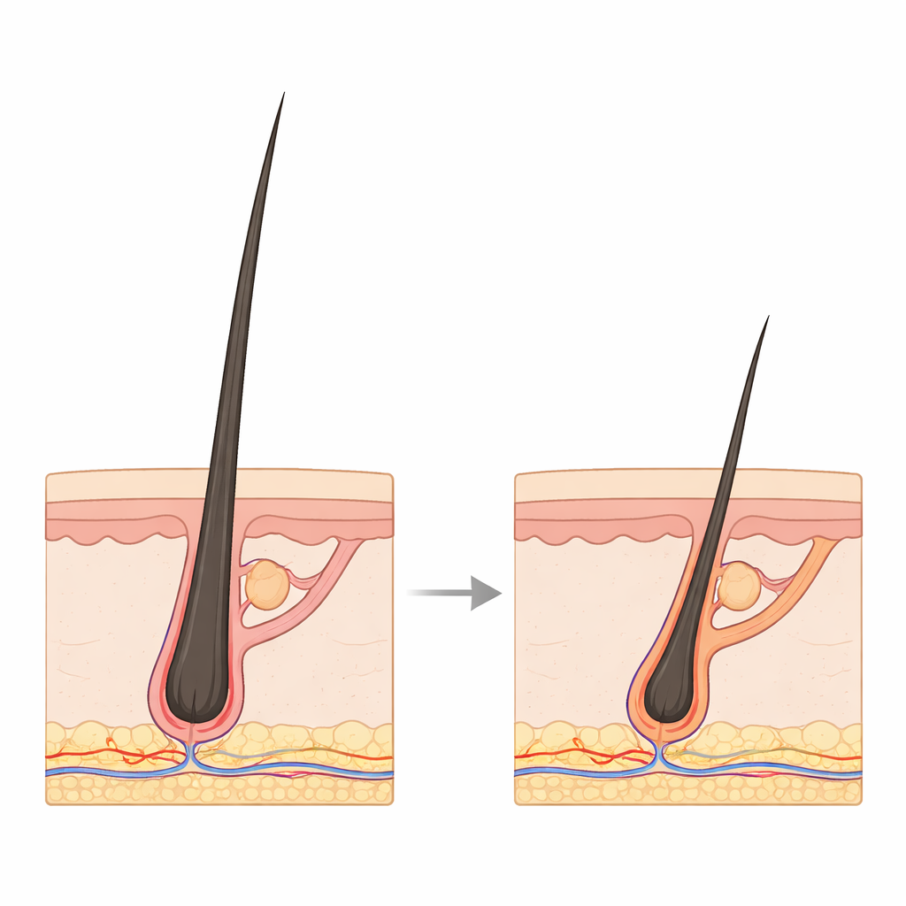
Each scalp hair is anchored in a miniature organ called a hair follicle, which repeatedly cycles through growth, regression, and rest. In androgenetic alopecia, or male pattern baldness, large follicles that produce thick hairs slowly shrink into much smaller units that only grow fine, barely visible hairs. Earlier work showed that stem cells at the base of these follicles largely remain, while their more active offspring—the progenitor cells that actually fuel hair production—gradually disappear, leaving a paradox: plenty of stem cells but too little hair. The authors tackled this puzzle by combining single-cell RNA sequencing, which reads out gene activity in thousands of individual cells, with spatial transcriptomics, which keeps track of where those cells sit inside intact scalp tissue.
Building a Cellular Map of the Balding Scalp
From balding and non-balding regions of the same men, as well as from healthy volunteers, the team isolated tens of thousands of cells from anagen (actively growing) follicles. They classified at least 20 major cell types, including stem cells, progenitor cells, hair-making matrix cells, immune cells, and several layers of surrounding support tissue. This high-resolution atlas showed that in balding follicles, key progenitor cells and matrix cells are reduced in number and display strong signs of inflammatory stress and cell death, closely linked to how much the follicle has shrunk. Meanwhile, immune cells that promote inflammation, particularly a subset called Th17 cells, were more abundant around these vulnerable follicles, hinting that low-grade inflammation and mechanical stress act together in hair loss.
The Follicle’s “Squeeze Sleeve” Misbehaves
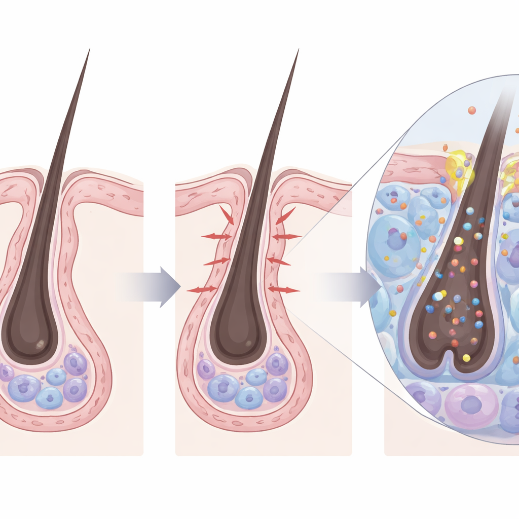
Wrapped around each human hair follicle is a multi-layered support structure called the connective tissue sheath. It contains smooth muscle–like cells, collagen fibers, and blood vessels and is closely related to a contractile layer previously shown in mice to help pull follicles upward during their normal regression phase. In balding human follicles, the genes that drive muscle contraction were strongly switched on in these sheath cells and nearby vessel wall cells. Microscopy confirmed that a molecular marker of contraction, phosphorylated myosin light chain, was higher in balding tissue. When the researchers stimulated this outer sheath with compounds that mimic body signals—such as a drug related to the nerve messenger acetylcholine, or the hormone dihydrotestosterone—they saw the sheath tighten, the follicles narrow, and hair growth slow in both cultured follicles and human scalp grafts on mice.
Mechanical Stress Kills Hair-Making Cells
The study then connected this physical squeezing to a specific “pressure sensor” in the follicle’s inner cells. Many of these cells, including stem and matrix cells, carry a mechanosensitive ion channel called PIEZO1 in their membranes. In balding follicles, PIEZO1 activity, reflected by rising calcium levels inside the cells, was elevated. Artificially activating PIEZO1 in cultured follicles reproduced the hallmarks of balding: slowed hair growth, increased death of progenitor cells, and reduced division of outer root sheath and matrix cells. Blocking PIEZO1, or relaxing the connective tissue sheath with a drug that inhibits a key contraction enzyme (ML-7), prevented these effects and rescued hair growth. The results suggest a chain of events: hormones and chemical signals drive the outer sheath to over-contract, that contraction mechanically activates PIEZO1 in hair-forming cells, and the resulting calcium surge nudges them toward death or dormancy.
Toward Gentler Treatments for Baldness
Finally, the team tested whether dialing down this squeezing could help human hair follicles already on the path to miniaturization. In cultured follicles from balding scalp areas, ML-7 eased contraction, reduced progenitor cell loss, and extended the growth phase, in some settings outperforming the standard topical drug minoxidil. In a humanized mouse model carrying transplanted human scalp, ML-7 improved hair growth from both non-balding and balding follicles and worked especially well when combined with minoxidil. To a lay reader, the key message is that male pattern baldness may not be due simply to “weak” hair roots, but to a stiff, overactive support sleeve that gradually strangles them. By targeting this mechanical problem—relaxing the sheath or blocking the pressure-sensing channel PIEZO1—future treatments could protect the hair-making cells and keep follicles larger and more productive for longer.
Citation: Li, G., Yang, L., Duan, S. et al. Single-cell transcriptomics reveals hair growth retardation mediated by aberrant connective tissue sheath contraction in male androgenetic alopecia. Nat Commun 17, 3252 (2026). https://doi.org/10.1038/s41467-026-70153-4
Keywords: androgenetic alopecia, hair follicle, mechanotransduction, PIEZO1, connective tissue sheath