Clear Sky Science · en
Site-specific profiling of structure and function of Igµ B cell receptor glycans
Why sugar decorations on immune cells matter
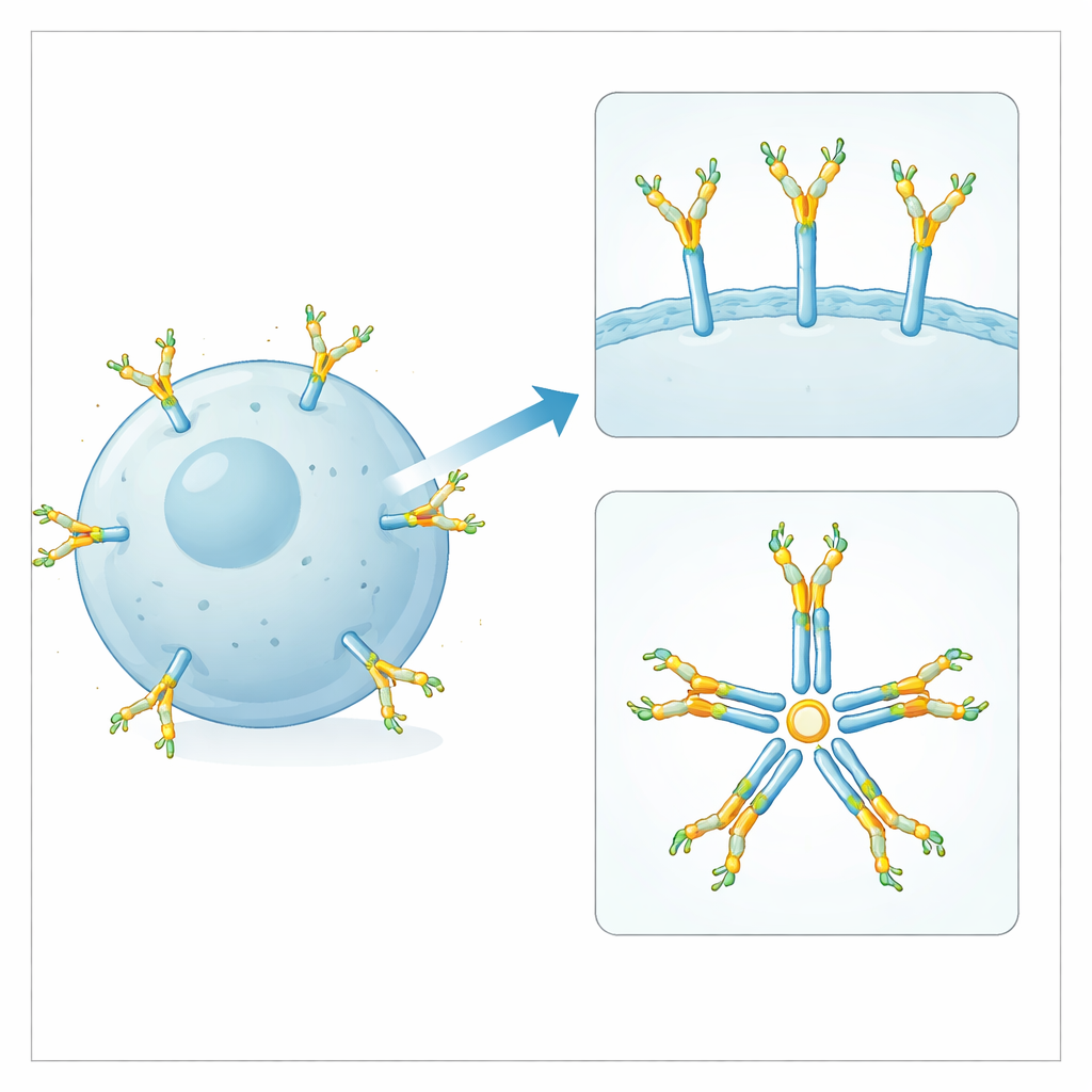
Our immune system’s B cells rely on antenna-like molecules, called B cell receptors, to sense threats and launch antibody responses. These receptors, and the antibodies they later secrete, are coated with tiny sugar trees known as glycans. Scientists know that such sugars can dramatically tune how antibodies behave, but have had far less insight into the sugars sitting directly on B cell receptors themselves. This study asks a simple but important question: do the sugar coats on a key human B cell receptor actually change how B cells work, or are they mainly there to support the antibodies that circulate in our blood?
Taking a closer look at a key B cell receptor
The researchers focused on the IgM-type B cell receptor, built from an Igµ heavy chain that carries four conserved glycan sites. These receptors sit in the membrane of naive B cells, which have not yet met their target, and memory B cells, which have. The team purified these receptors from human blood, then used high-resolution mass spectrometry to measure which kinds of glycans occupy each site. They compared naive and memory cell receptors with IgM antibodies from the same donors’ blood serum, which are released versions of the same basic protein scaffold. This allowed them to ask both whether maturation of B cells reshapes receptor glycans and how membrane-bound receptors differ from free-floating IgM antibodies.
Surprising stability across the life of a B cell
The detailed profiling showed that naive and memory IgM B cell receptors carry almost identical collections of glycans at all four positions. Three sites (on the first three constant domains) mainly bear complex, branched glycans that are fucosylated, galactosylated and heavily capped with sialic acid, while the fourth site largely holds simpler, mannose-rich structures. Moving from a naive to a memory state therefore does not involve a visible remodeling of these sugar coats. In other words, the chemical decorations on this receptor appear to be a stable feature of the molecule, rather than a switch that B cells flip as they learn from prior infections.
How membrane receptors differ from free antibodies
When the team compared B cell receptors to IgM antibodies in serum, patterns did emerge. On the three complex glycan sites, the membrane receptors carried more sialic acids and fewer so-called bisecting sugar branches than their antibody counterparts. At the mannose-rich site, the receptors retained more mannose residues than serum IgM. Using engineered B cell lines that expressed the same IgM either as a receptor or as a secreted antibody, the researchers saw the same trends. This strongly suggests that the differences are built into the molecular context: membrane-anchored, single-unit receptors are processed differently by the cell’s glycan-adding enzymes than are pentameric, secreted IgM antibodies with an extra tailpiece.
Testing whether specific sugars change B cell behavior
To learn whether any one glycan site directly alters B cell behavior, the scientists created cell lines with precise mutations that removed individual glycan sites from the IgM receptor. They then measured how well these mutant receptors reached the cell surface, bound their target antigens, were pulled inside the cell after stimulation, and triggered early signaling through a key enzyme called Syk. Removing any single glycan did not stop receptors from being displayed on the surface or from being internalized or signaling once triggered. However, loss of the glycan at one particular site, called N209, consistently reduced how strongly the receptor could grab onto its antigen, across several different antigen specificities.
How one sugar site supports receptor structure
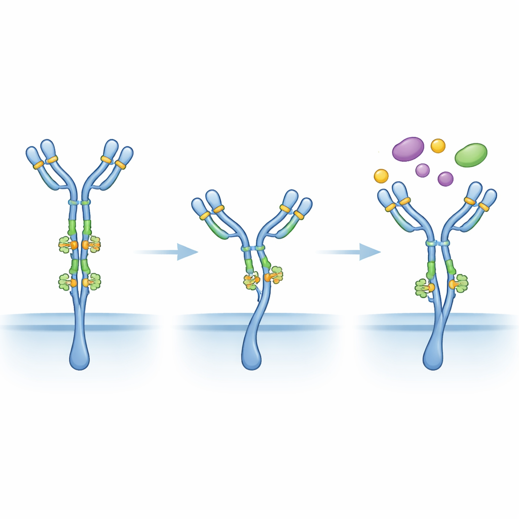
To understand why the N209 site mattered, the team modeled the three-dimensional structure of the IgM receptor using cryo-electron microscopy data combined with computer simulations of the attached glycans. These simulations revealed that the sugars at N209 form a flexible, space-filling cluster where the two halves of the receptor meet. This cluster behaves like a dynamic spacer, helping keep the two antigen-binding arms extended away from the receptor’s base and the cell membrane. When this glycan is missing, the model suggests the receptor can collapse into a less favorable shape for catching antigen, even though its ability to be internalized and to signal once triggered remains intact.
What this means for immunity and antibodies
Overall, the work shows that the sugar coats on IgM B cell receptors are highly conserved between naive and memory B cells and, with one notable exception, do not strongly control core receptor functions like expression, internalization or early signaling. The N209 sugar seems to act more like a structural brace, supporting optimal antigen binding, than as a general on–off switch for B cell activity. The clear differences between receptor glycans and those on circulating IgM antibodies, combined with previous findings that IgM glycans shape how antibodies activate complement and other effector pathways, suggest that these sugar decorations evolved primarily to fine-tune antibody behavior in the bloodstream. The sugars on the receptor itself appear to be passengers rather than drivers of B cell function, with the key structural N209 site as an elegant exception.
Citation: Holborough-Kerkvliet, M.D., Hafkenscheid, L., Kroos, S. et al. Site-specific profiling of structure and function of Igµ B cell receptor glycans. Nat Commun 17, 3507 (2026). https://doi.org/10.1038/s41467-026-70121-y
Keywords: B cell receptor glycosylation, IgM antibodies, glycans, humoral immunity, antibody structure