Clear Sky Science · en
Swapped and non-swapped TRAAK states co-exist in membranes at a ratio influenced by temperature
Why this tiny gate in nerve cells matters
Every thought, touch, and heartbeat depends on electrical signals flowing through our cells. A key player in shaping those signals is a family of protein "leak" channels that let potassium ions slip across cell membranes. One member, called TRAAK, helps set how excitable nerve cells are and even participates in sensing temperature and mechanical force. This study uncovers how a small movable "cap" on top of TRAAK behaves in real cell membranes, how heat shifts its preferred shape, and how the surrounding fats in the membrane form a special neighborhood that tunes the channel’s function. 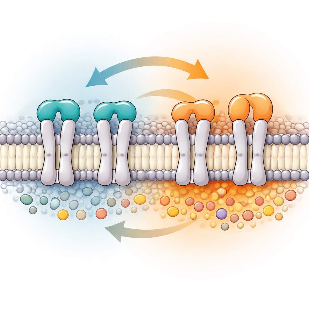
Two ways to arrange a microscopic gate
TRAAK belongs to a group of so‑called two‑pore domain potassium channels that act as background leak pathways, keeping the electrical balance of many cell types, including those in the brain and heart. Unlike more familiar four‑part potassium channels, TRAAK is built from two large subunits and carries a distinctive cap that juts above the membrane and splits the route that ions take. Earlier high‑resolution structures revealed that this cap can adopt two arrangements: a "swapped" layout in which parts of one subunit cross over to contact the other, and a "non‑swapped" layout in which each subunit’s pieces stay with their original partner. Until now, no one knew whether both forms actually coexist in natural membranes, how common each form is, or what tips the balance between them.
Watching moving parts with magnetic rulers
To tackle this, the authors used a specialized magnetic technique called pulse dipolar electron paramagnetic resonance, which can measure distances of a few billionths of a meter between tiny magnetic tags. They engineered human TRAAK so that only one of the two subunits carried a pair of these tags at carefully chosen positions bridging the cap and the membrane‑spanning region. By doing so, they could distinguish the two cap shapes because each produces a different tag‑to‑tag distance, much like measuring the span between two hinges in alternative door designs. They also embedded TRAAK in membrane fragments held together without harsh detergents, preserving the nearby natural lipids and ensuring that the full‑length protein—complete with all regulatory parts—remained functional. 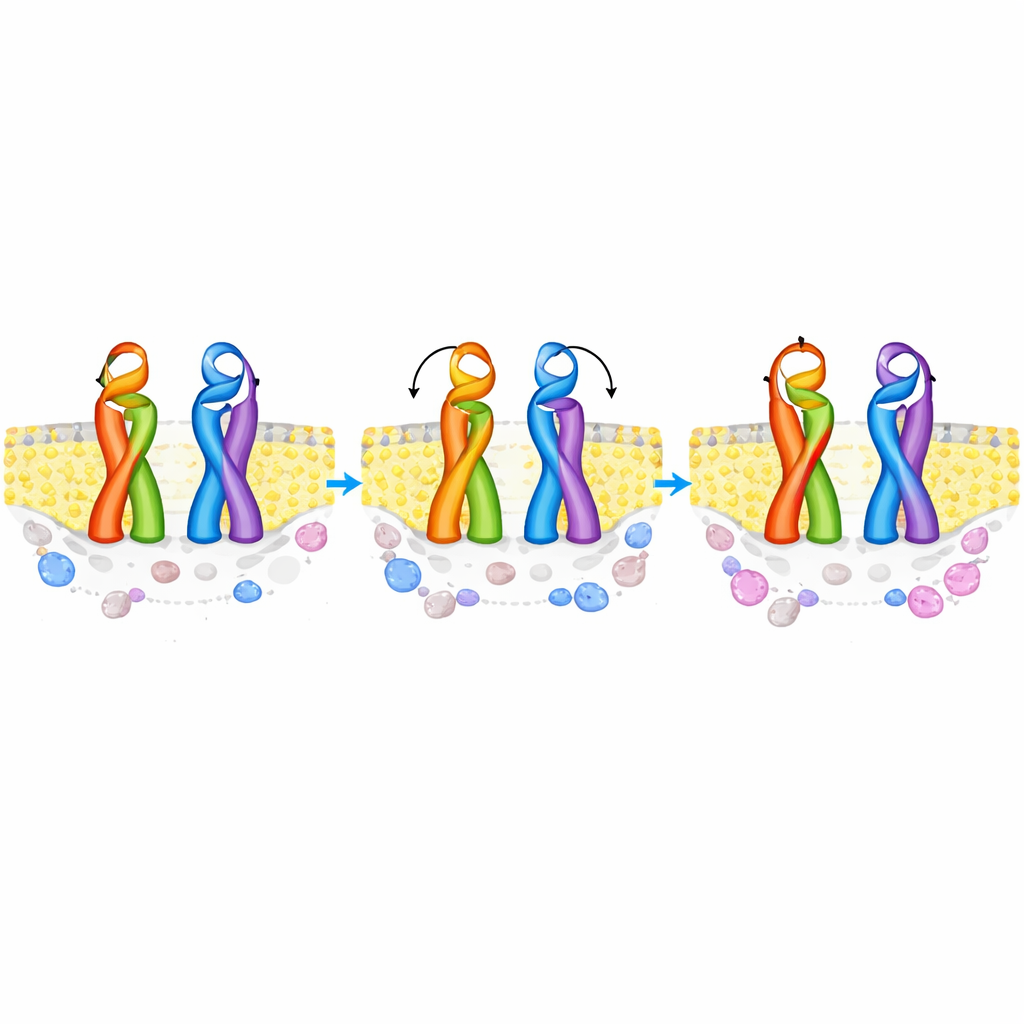
Two shapes share the stage, and heat changes the script
The distance measurements revealed two distinct populations that matched computer predictions for the swapped and non‑swapped caps, proving that both forms coexist in the same membrane ensemble. At a near‑room temperature of 19 °C, the swapped form made up a modest majority of the channels, with the non‑swapped form still representing a substantial minority. When the researchers prepared the membrane samples at a warmer 40 °C, the balance shifted further toward the swapped state, and the non‑swapped configuration became rarer. Computer simulations of TRAAK in model membranes suggested that the swapped arrangement is inherently more stable and less sensitive to temperature, whereas the non‑swapped form becomes energetically less comfortable as the membrane and its lipids reorganize with heat.
A custom lipid neighborhood around TRAAK
Beyond shape‑shifting, TRAAK also proved selective about the company it keeps in the membrane. By analyzing the fats that stayed bound to TRAAK during detergent‑free purification, the team found a strong enrichment of phosphatidylinositol and its signaling variants—lipids known to influence many ion channels—along with other negatively charged species. Surprisingly, the most common membrane lipid, phosphatidylcholine, was essentially absent from TRAAK’s immediate surroundings, even though it dominates the host cell’s membrane. When reconstituted into artificial vesicles, some of the enriched lipids activated TRAAK currents, while others dampened them or had little effect, showing that the channel’s preferred lipid microdomain is not just structural but also functionally important.
What this means for brain signals and beyond
Together, these findings show that TRAAK does not exist in a single frozen shape. Instead, it shifts between swapped and non‑swapped cap states whose relative abundance depends on temperature and on the fine details of the surrounding membrane. The swapped state is more common and becomes even more favored as things warm up, hinting that cap rearrangements may contribute to how TRAAK responds to heat and mechanical cues in living cells. By preserving native lipids and precisely reading out rare conformations, the authors demonstrate a powerful way to link microscopic shape changes to the special lipid pockets that host them. This framework can now be applied to other leak channels that influence pain, anesthesia, and neurological disease, helping to explain how subtle alterations in membrane composition and protein shape can reshape the electrical language of our cells.
Citation: Ma, Y., Ackermann, K., Waheed, Q. et al. Swapped and non-swapped TRAAK states co-exist in membranes at a ratio influenced by temperature. Nat Commun 17, 3522 (2026). https://doi.org/10.1038/s41467-026-70027-9
Keywords: TRAAK channel, potassium leak channels, membrane lipids, temperature sensing, ion channel structure