Clear Sky Science · en
Quantitative phase gradient microscopy with spatially entangled photons
Seeing the Invisible in Gentle Detail
Many of the most important samples in biology and materials science—like living cells, tissue slices, or thin films—are nearly transparent. They barely absorb light, so they look like faint shadows in an ordinary microscope. The study described here introduces a new way to turn those almost invisible variations into crisp, measurable pictures of both the shape and internal thickness of such samples, while using extremely little light. This makes it especially attractive for examining fragile, light‑sensitive specimens.
From Blurry Outlines to Precise Maps
Classic phase‑contrast microscopes, invented in the 1930s, changed biology by turning tiny changes in how light slows down inside a cell into visible contrast. But they mostly give a qualitative view—good for seeing structure, poor for measuring exact thickness or refractive index. Modern “quantitative phase imaging” methods try to turn those subtle delays into precise height maps with nanometer‑level sensitivity. However, they usually rely on complex interferometers, moving parts, arrays of microlenses, or heavy computer processing with many images. These requirements can make the systems bulky, delicate, slow, and sensitive to environmental noise.
Entangled Light as a New Kind of Probe
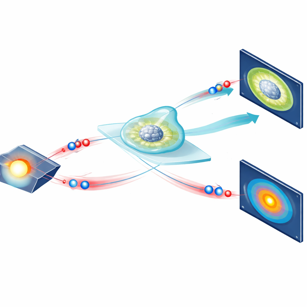
The authors propose and demonstrate a different route that uses quantum light—specifically, pairs of spatially entangled photons. These pairs are born together in a special crystal and are tightly linked: their positions are strongly correlated, and their directions of travel are strongly anti‑correlated. In the new microscope, both photons in each pair pass through the transparent sample, but they are observed in two different ways. One camera records where one photon lands in a sharp “near‑field” image, while another camera captures the partner photon in the “far field,” where its position reveals subtle changes in direction. By looking only at photons that arrive as true pairs, and by analyzing their joint pattern, the system retrieves both the sample’s brightness and how its thickness varies across the field of view, all without using an interferometer or scanning.
Turning Tiny Bends of Light into Height Maps
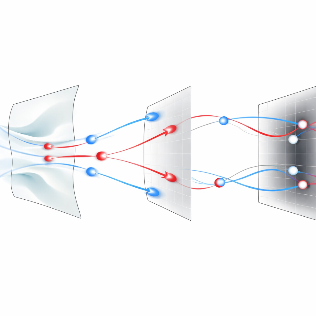
When light passes through a region of a sample where the optical thickness changes gradually, its wavefront tilts slightly, like water flowing over a gentle underwater hill. In this method, such local slopes show up as small shifts in where the partner photon lands in the far‑field camera. For each pixel in the near‑field image, the researchers calculate the average shift of the correlated spots in the far‑field image; this shift is directly linked to the local “phase gradient,” or how fast the optical thickness is changing at that point. A mathematical reconstruction then stitches all these local gradients together into a full map of phase, which can be read as an effective thickness map of the object. Using standard test patterns, the team shows that they can resolve features as small as 2.76 micrometers and accurately measure phase steps as tiny as about one‑hundredth of the light’s wavelength, all while illuminating the sample with only about one hundred femtowatts of power—billions of times weaker than a typical laser pointer.
Seeing Clearly Through a Noisy Glow
Real‑world imaging often suffers from messy, changing background light, such as glow from fluorescent markers or other stray sources. Conventional phase‑gradient methods can be badly distorted by such backgrounds and typically require extra steps to measure or filter them out. Here, the built‑in timing correlations of entangled photon pairs become a powerful filter. The camera records the arrival time of every detected photon, and only those pairs that arrive within a tight time window are treated as coming from the entangled source. By also measuring “accidental” coincidences in a shifted time window—where no real pairs should occur—the researchers can estimate the contribution from random background light and subtract it. They show that this correction recovers much more accurate phase images even when a strong, moving background beam is deliberately added to the system.
New Possibilities for Gentle, Precise Imaging
The work delivers a proof‑of‑concept microscope—called quantum correlation phase gradient microscopy—that gives precise, quantitative images of transparent samples without interferometers, moving parts, or iterative guess‑and‑refine algorithms. It works at extremely low light levels, making it promising for studies of sensitive biological samples, and it naturally resists complex, time‑varying background light. Beyond imaging living cells, the authors envision applications in fine‑tuning optical systems, probing delicate materials, and eventually extending the approach to three‑dimensional imaging as detector technology improves.
Citation: Zhang, Y., Moreau, PA., England, D. et al. Quantitative phase gradient microscopy with spatially entangled photons. Nat Commun 17, 3108 (2026). https://doi.org/10.1038/s41467-026-69881-4
Keywords: quantum imaging, entangled photons, phase microscopy, low-light imaging, adaptive optics