Clear Sky Science · en
Pattern-enhanced Resonant Soft X-ray Scattering for Operando monitoring of electrochemical solid-liquid interfaces
Why tiny buried surfaces matter
Many of today’s most important technologies, from rechargeable batteries to devices that make hydrogen fuel from water, rely on what happens where solids touch liquids. These thin boundary layers control how fast reactions run and how long devices last, but they are extremely hard to watch in action: they are buried beneath liquids, only a few atoms thick, and constantly changing. This study introduces a new X‑ray–based method that can track both the structure and chemistry of such hidden interfaces in real time without damaging them.
Turning the sample into part of the microscope
The authors develop a technique called Pattern‑enhanced Resonant Soft X‑ray Scattering, or PE‑RSoXS. Instead of treating the sample as a passive object, they deliberately sculpt it into a precisely spaced “line‑grating” of metal nanostripes. When soft X‑rays pass through this patterned surface, the nanostripes act like tiny optical components that bend and interfere with the X‑rays in a controlled way. By tuning the X‑ray energy to match how specific elements absorb light, the method becomes sensitive not only to shape and thickness, but also to the chemical state of the atoms at the surface. The repeated pattern makes the scattered X‑ray signal add up coherently, boosting its strength by several orders of magnitude compared with a single, unpatterned feature. 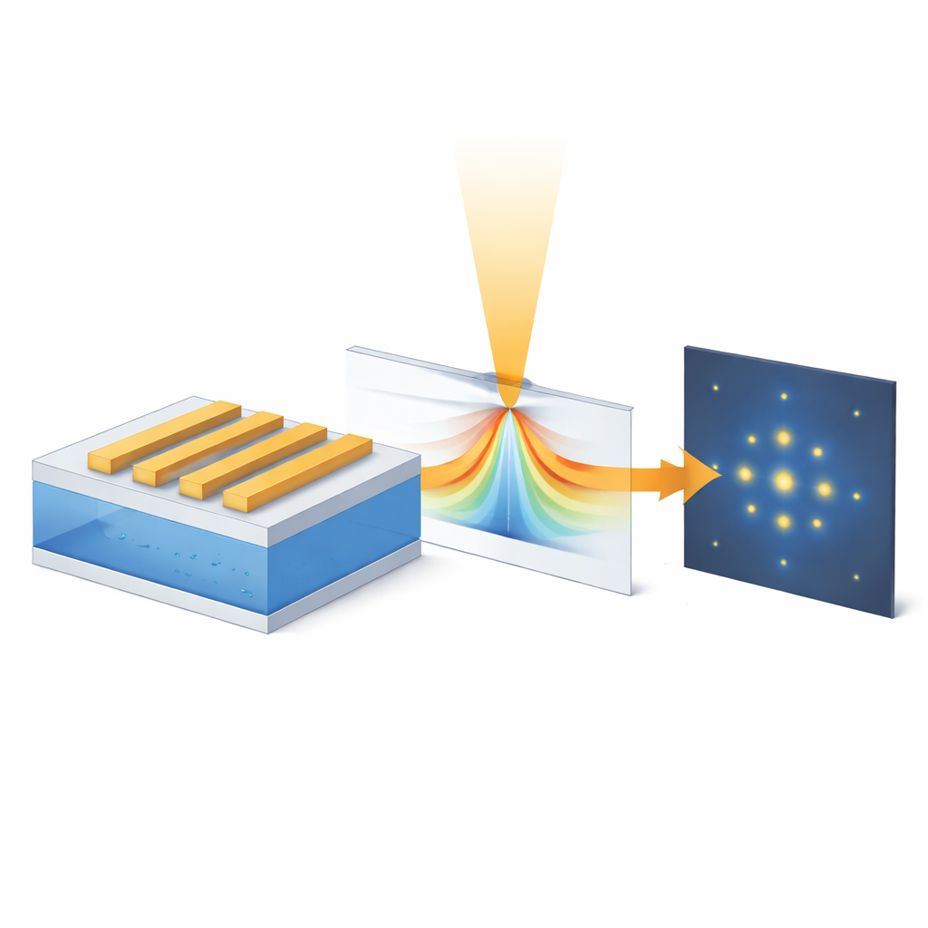
Watching a working water‑splitting electrode
To showcase PE‑RSoXS, the team studies nickel electrodes that drive the oxygen‑evolution reaction, a key step in splitting water to make green hydrogen. They fabricate nickel nanostripes about 100 nanometers wide and 50 nanometers tall on thin windows that can be mounted in a tiny flow cell filled with alkaline solution. While the electrode operates at different voltages, soft X‑rays tuned near the nickel absorption edge illuminate the stripes, and a detector records the resulting pattern of bright diffraction spots. Because different diffraction orders respond differently to changes in the outer shell versus the inner core of each stripe, the researchers can separate signals from the buried interface from those of the bulk metal underneath.
Revealing sub‑nanometer changes and active states
By comparing the measured scattering patterns with detailed computer simulations of how X‑rays propagate through core‑shell stripes, the authors reconstruct how the nickel surface evolves under operating conditions. At open circuit and at a moderate voltage, the outer shell of oxidized nickel remains thin, and the overall stripe width even contracts slightly, suggesting the formation of a denser surface layer. When the voltage is raised into the regime where oxygen is actively produced, the shell thickens by only a couple of nanometers and the stripes swell modestly in width—changes far below the X‑ray diffraction limit but still detectable through their influence on the diffraction intensities. At the same time, the energy‑dependent scattering signals reveal that nickel atoms in the shell shift from a lower oxidation state to a higher one associated with the most active form of the catalyst.
Probing dynamics that other tools miss
The method is fast and gentle: each scattering pattern can be taken in a millisecond with extremely low X‑ray dose, avoiding damage that can plague electron‑microscope studies. Because hundreds of identical nanostripes contribute to the signal, the measurements are statistically robust rather than tied to a single tiny region. Additional simulations show that PE‑RSoXS is sensitive not only to shell thickness and composition, but also to where an oxidized layer sits within each repeating unit, hinting at a practical spatial resolution better than a nanometer in terms of resolving interfacial structure.
How this advances clean‑energy research
In everyday terms, this work turns a catalytic surface into its own finely tuned antenna for X‑rays, allowing researchers to “listen in” on how buried interfaces rearrange and change their chemistry while a reaction runs. The authors demonstrate that PE‑RSoXS can pinpoint when and where the catalytically active nickel phase forms, and how much the material swells, all under realistic liquid conditions. Because the approach can be adapted to other elements by retuning the X‑ray energy and redesigning the patterns, it offers a versatile way to study a broad range of energy and catalytic systems. Ultimately, such insights can guide the design of longer‑lasting batteries, more efficient fuel‑forming electrodes, and other technologies that depend on fragile, hidden interfaces.
Citation: Li, H., Andrle, K., Zhang, Q. et al. Pattern-enhanced Resonant Soft X-ray Scattering for Operando monitoring of electrochemical solid-liquid interfaces. Nat Commun 17, 2997 (2026). https://doi.org/10.1038/s41467-026-69852-9
Keywords: electrochemical interfaces, soft X-ray scattering, water splitting, nickel catalysts, operando characterization