Clear Sky Science · en
Structural and dynamic insights into agonist recognition and function of the thromboxane A2 receptor
Why this blood-clotting gatekeeper matters
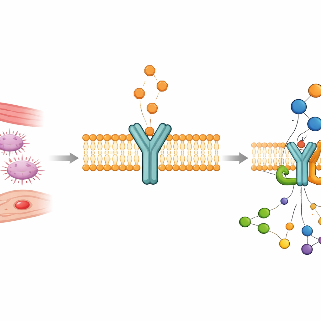
The body keeps blood flowing smoothly while still being able to plug a wound in seconds. A key player in this balancing act is the thromboxane A2 receptor, a tiny protein switch on platelets and smooth muscle cells that helps control clotting, vessel narrowing, and inflammation. When this switch misfires or is overactive, it can contribute to heart attacks, strokes, lung disease, fibrosis, and even some cancers. This study reveals, in unprecedented atomic detail, how this receptor recognizes its activating molecules and turns on inside the cell—knowledge that could guide safer, more precise drugs to fine‑tune clotting and vascular tone.
A short‑lived signal with long‑lasting impact
Thromboxane A2 is a fat‑derived messenger produced in platelets and blood vessel walls. It is chemically fragile, breaking down in about half a minute, yet in that brief window it tells platelets to clump and causes the surrounding muscle to contract. It does this by binding to the thromboxane A2 receptor, a member of the large family of G protein‑coupled receptors that sit in the cell membrane and relay signals inward. Because natural thromboxane disappears so quickly, it has been hard to capture exactly how it docks to its receptor and flips it into the active, signalling state. The researchers sidestepped this limitation by using two stable, high‑potency look‑alikes, U46619 and I‑BOP, which mimic thromboxane but last long enough to be studied structurally.
Seeing the receptor in action
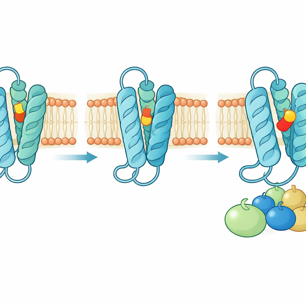
Using cryogenic electron microscopy, the team imaged the human receptor bound to each of these synthetic agonists while it was coupled to its main intracellular partner, the Gq protein. The resulting three‑dimensional structures, sharpened to near‑atomic resolution, show the receptor spanning the cell membrane with seven tightly packed helices. The agonist molecules sit deep inside this bundle in an L‑shaped pose, protected from water that would otherwise degrade them. On the inside of the cell, one of the helices swings outward, opening a cradle into which the G protein’s tail fits, priming the cell for a surge of downstream calcium and enzyme activity. Biochemical signalling assays confirmed that these complexes faithfully represent the active, working state of the receptor.
A hidden doorway through the membrane
The structures, together with extensive computer simulations, indicate that thromboxane‑like molecules do not primarily approach the receptor from the watery outside of the cell. Instead, they slip in from within the fatty membrane itself. A movable gap between two of the outer helices, aided by flexible aromatic side chains and nearby cholesterol molecules, acts as a molecular gate. In the inactive state this gap is open, allowing entry; once the agonist is buried in the pocket, the gate swings shut, sealing the ligand in place. Mutating key residues that form this gate, or altering their interaction with cholesterol, severely disrupts receptor signalling—and helps explain rare human bleeding disorders caused by naturally occurring mutations at these sites.
An unconventional way to flip the switch
Most related receptors are activated when a ligand nudges a conserved “toggle” amino acid that then drives a large movement of one of the helices. In the thromboxane receptor, that classic toggle is present but plays a supporting role. Instead, the study finds that a nearby glutamine and asparagine pair on the inner side of the ligand pocket acts as the real switch. Agonists donate a hydrogen bond in a way that forces these residues to rearrange their connections, pulling one helix inward, letting another rotate, and finally freeing the outward‑moving helix that creates space for the G protein. Antagonist drugs, by contrast, sit higher in the pocket and bond differently to the same glutamine, stabilizing an inactive network of contacts that keeps the G protein site closed. Dozens of carefully chosen mutations, tested in living cells, support this revised view of how the receptor turns on and off.
From atomic blueprints to better medicines
Overall, the work provides a full mechanistic picture of how unstable thromboxane signals are captured from the membrane, locked into a sheltered cavity, and converted into a robust intracellular response via an unusual activation switch. By explaining why some ligands strongly activate the receptor while others block it, and by mapping how disease‑linked mutations derail these processes, the study offers a detailed blueprint for next‑generation drugs. Such compounds could more precisely dampen excessive clotting, vessel constriction, and fibrosis—or selectively block harmful thromboxane signalling in cancer—while preserving the receptor’s vital role in everyday vascular health.
Citation: Krawinski, P., Matzov, D., Ryder, A. et al. Structural and dynamic insights into agonist recognition and function of the thromboxane A2 receptor. Nat Commun 17, 3071 (2026). https://doi.org/10.1038/s41467-026-69844-9
Keywords: thromboxane receptor, blood clotting, G protein–coupled receptor, cryo-EM structure, cardiovascular disease