Clear Sky Science · en
Cell-specific responses of Anopheles gambiae fat body to blood feeding and infection at single-nuclei resolution
Why mosquito body fat matters
Malaria-carrying mosquitoes do far more with their “body fat” than simply store energy. In Anopheles gambiae, a major African malaria vector, this soft tissue helps power egg production and acts as a front-line immune organ. The study described here builds a detailed cellular atlas of this tissue and shows how its different cells react when a mosquito takes a blood meal or encounters infection—insights that could ultimately inform new ways to curb malaria transmission.
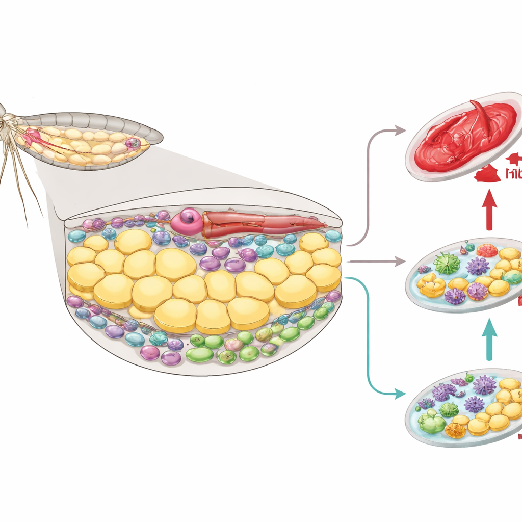
A hidden organ with many jobs
The mosquito fat body lines much of the abdomen and behaves like a cross between the human liver and body fat. It stores sugars and lipids, makes proteins that circulate in the insect’s blood-like fluid, and helps detoxify chemicals. It also produces molecules that fight invading microbes and supports egg development after a blood meal by supplying yolk and lipids to the ovaries. Yet, despite these vital roles, the fine-scale organization of this tissue—what cell types it holds and which jobs each cell type performs—has remained unclear.
Mapping thousands of tiny nuclei
To tackle this, the researchers isolated almost 100,000 nuclei from the abdominal body wall of female mosquitoes and read out which genes were active in each nucleus. This “single-nucleus RNA sequencing” approach sidesteps the challenge of breaking apart very fatty, fragile cells. By grouping nuclei with similar gene activity patterns, the team identified 12 clusters that corresponded to seven main cell types. Most cells (about 85%) were fat body trophocytes, the workhorse cells that stockpile nutrients. Other clusters represented immune cells stuck to or embedded in the tissue, cuticle-forming epidermal cells, nerve cells, pericardial cells near the heart, and specialized oenocytes involved in lipid chemistry.
Different cells, different specialties
Even within the trophocytes, the team found five subgroups with distinct roles. Two “basal” groups appeared to handle routine metabolism. A third subgroup showed elevated activity in energy and protein production pathways, hinting at a metabolic specialist role. A fourth subgroup stood out for its constant expression of immune-related genes, suggesting a built-in surveillance function. A fifth group appeared only after a blood meal and strongly activated genes for yolk proteins and related processing enzymes, marking these cells as central players in egg provisioning. Under the microscope, messages for the main yolk protein concentrated in the layer of trophocytes facing the mosquito’s blood and even piled up at the side of each cell that touches this fluid, consistent with directed export toward the ovaries.
How blood meals and infections reshape the tissue
The team then compared mosquitoes that were sugar-fed, blood-fed, injected with bacteria, or infected with malaria parasites that trigger a long-lasting immune “priming.” After a blood meal, many trophocytes flipped from the basal to the vitellogenic state, turning on genes for yolk and lipid export while dialing down genes involved in basic carbohydrate metabolism and some immune functions. Markers of DNA replication surged, and most trophocyte nuclei incorporated a synthetic building block of DNA without showing signs of cell division—evidence that these cells increase their DNA content to boost production capacity rather than multiply in number. In contrast, bacterial infection led to a strong, rapid immune response: antimicrobial peptides and other defense genes rose sharply across trophocytes, immune cells, epidermal cells, and pericardial cells. One trophocyte subgroup with pre-activated immune genes responded especially strongly, while other subgroups adjusted energy pathways, pointing to a trade-off between defense and metabolism.
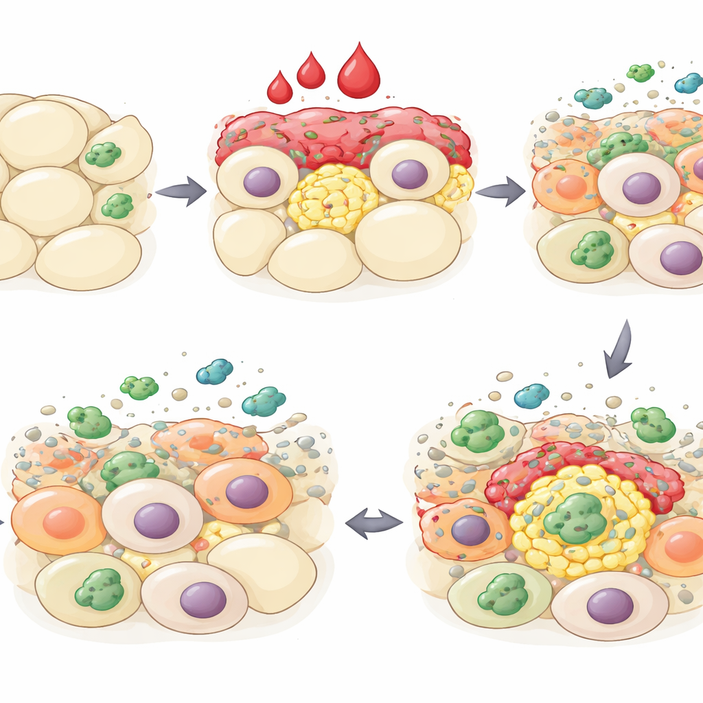
Primed defenses for repeat encounters
When mosquitoes were exposed to malaria parasites in a way known to create lasting immune readiness, the biggest change occurred in oenocytes. These cells upregulated many enzymes involved in fatty acid and hydrocarbon production, including components tied to the synthesis of lipid-based signaling molecules that help establish immune memory. Nearby immune cells adjusted genes related to adhesion and cholesterol use, consistent with closer interaction with the fat body and possible production of additional bioactive lipids. Together, these shifts suggest that the tissue’s lipid factories and immune cells work together to heighten preparedness for future infections.
What this means for malaria control
Overall, the study reveals the mosquito fat body as a highly organized and flexible organ where distinct cell types and subtypes coordinate metabolism, reproduction, and immunity in space and time. Blood feeding temporarily reprograms many trophocytes into egg-support specialists that amplify their DNA to meet intense production demands, while infection activates a shared but cell-type-tailored defense program. Oenocytes emerge as key players in long-term immune priming. By charting this complexity at single-cell resolution, the work provides a blueprint for targeting specific cell states or processes—such as yolk production or immune memory—to reduce mosquito fertility or their ability to host and transmit malaria parasites.
Citation: de Carvalho, S.S., McNinch, C., Barletta, AB.F. et al. Cell-specific responses of Anopheles gambiae fat body to blood feeding and infection at single-nuclei resolution. Nat Commun 17, 3119 (2026). https://doi.org/10.1038/s41467-026-69806-1
Keywords: mosquito immunity, fat body cells, single nucleus RNA sequencing, blood feeding, malaria vector biology