Clear Sky Science · en
Villification of the intestinal epithelium is driven by Foxl1 through activation of PDGFRα and BMPs
How the Gut Gets Its Inner Landscape
The lining of your small intestine looks a bit like a dense underwater forest, full of tiny finger-like projections called villi. These structures massively increase the surface area available to absorb nutrients from food. This paper uncovers how, before birth, a smooth fetal gut tube reshapes itself into this intricate landscape, and pinpoints a key cell type and gene—Foxl1—that help organize this transformation from the tissue just beneath the intestinal lining.
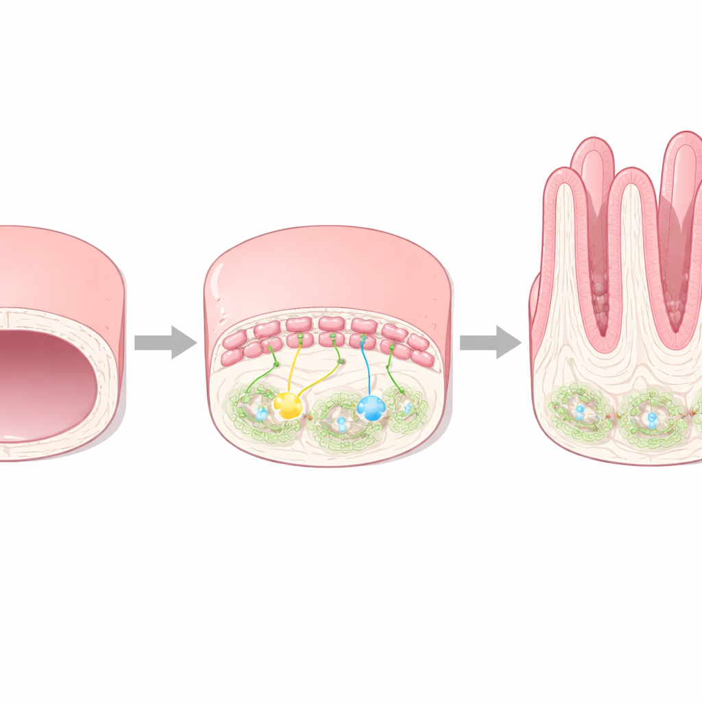
From Smooth Tube to Textured Surface
Early in mammalian development, the gut is a simple, smooth cylinder made of an inner sheet of epithelial cells wrapped by supporting tissue called mesenchyme. As the fetus grows, this inner surface must transform into a series of ridges and villi to handle future digestion. The authors show that this remodeling, known as villification, depends on a specific group of mesenchymal cells lying just under the epithelium. These cells respond to signals coming from the epithelial layer and in turn send instructions back, helping the lining first bend, then separate into dividing zones at the base and more mature zones at the tips of future villi.
A Supporting Cell Type with a Directing Role
The study focuses on Foxl1, a gene active in a specialized population of stromal cells called telocyte progenitors that hug the underside of the gut epithelium. Using fluorescent reporter mice and single-cell RNA sequencing, the researchers discovered that these Foxl1-positive cells are not all the same. They divide into two related subgroups: one group sits beneath regions destined to become villus tips, while the other lies next to future intervillus areas where stem and progenitor cells will reside. The villus-associated subgroup strongly expresses molecules such as PDGFRα and several BMP family proteins, which are known to influence how tissues grow, fold, and quiet down their cell division.
What Happens When the Director Goes Missing
To test how important Foxl1 is, the team examined mice lacking this gene. These animals formed fewer and later villi during fetal stages, and their small intestines showed long ridges rather than regularly spaced emerging villi. Microscopy and molecular analysis revealed that, although Foxl1-positive telocytes were still present in number, they lost much of their PDGFRα and BMP activity. As a result, the normal BMP signal that should restrain cell division and help define villus versus intervillus territories was weakened. Markers of proliferation and of a growth-promoting pathway spread into regions that should already be maturing, and the pattern of epithelial folds that ordinarily seed villi was disrupted. Some surface cells that lost proper contact with underlying tissue went on to die, underscoring how mechanical organization and signaling are tightly linked.
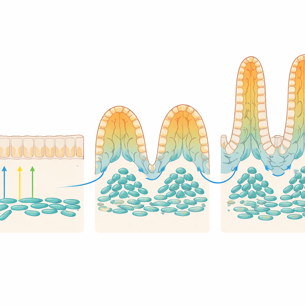
Fine-Tuning Tissue Orientation and Cell Types
Beyond growth control, Foxl1-positive telocytes also influenced how cells align and specialize. The authors found that these cells help activate a planar cell polarity program—signals that tell cells which way is “across” the tissue plane—through a gene called Fat4. Without Foxl1, expression of Fat4 in the villus-associated telocyte group dropped, nearby stromal cells failed to reorient along forming villi, and the characteristic boundary folds that mark future villus edges appeared less often. At the same time, the epithelial population shifted away from secretory progenitor cells—which will later produce mucus and hormones—toward more undifferentiated, dividing cells. Markers of early secretory cells, including future mucus-producing goblet cells, were transiently reduced in Foxl1-deficient intestines.
Why This Matters for a Healthy Gut
Together, these findings paint Foxl1-positive telocytes as local conductors that translate incoming developmental signals into a coordinated program: they boost PDGFRα and BMP pathways to shape where villi rise and where cells keep dividing, and they activate polarity cues to align the tissue architecture. When Foxl1 is removed, villus formation is delayed, boundaries blur, and specialized secretory cells are late to appear, though other factors eventually compensate before birth. For a lay reader, the key message is that a thin layer of supporting cells just beneath the intestinal lining quietly choreographs how the gut’s inner surface becomes a highly folded, efficient absorptive organ—an essential step for life after birth.
Citation: Zhu, G., Rozenberg, G., Lahori, D. et al. Villification of the intestinal epithelium is driven by Foxl1 through activation of PDGFRα and BMPs. Nat Commun 17, 3122 (2026). https://doi.org/10.1038/s41467-026-69791-5
Keywords: intestinal villi, gut development, stromal signaling, Foxl1 telocytes, fetal intestine