Clear Sky Science · en
Structural organization of HBV pgRNA genome driven by phase separation in capsid confinement
How a Tiny Virus Packs a Big Genome
Hepatitis B virus (HBV) is a major cause of liver disease worldwide, yet its genetic material fits inside a protein shell only about a hundred billionths of a meter across. This paper explores a basic but long-standing puzzle: how can the virus squeeze its RNA genome into such a cramped space while still keeping it mobile enough to copy itself? Using computer simulations and lab experiments, the authors uncover a physical process, similar to how oil droplets form in water, that lets HBV neatly organize its genome inside its shell and may offer new ways to disrupt the virus.
A Crowded World Inside the Viral Shell
Inside HBV’s protein shell, or capsid, lies pregenomic RNA (pgRNA), a long single-stranded molecule that serves as the template for making viral DNA. The inner surface of the shell bristles with flexible, positively charged protein tails that are attracted to the negatively charged RNA. Detailed atomic simulations show that, rather than forming a solid lump in the center, the pgRNA quickly moves to the inner wall and forms a hollow shell-like layer that closely hugs the capsid. Within this layer, dense patches of RNA and protein tails coexist with more open, porous regions. On average, this arrangement matches the highly symmetric patterns seen in cryo-electron microscopy images, but each individual virus particle can look quite different at any given moment. 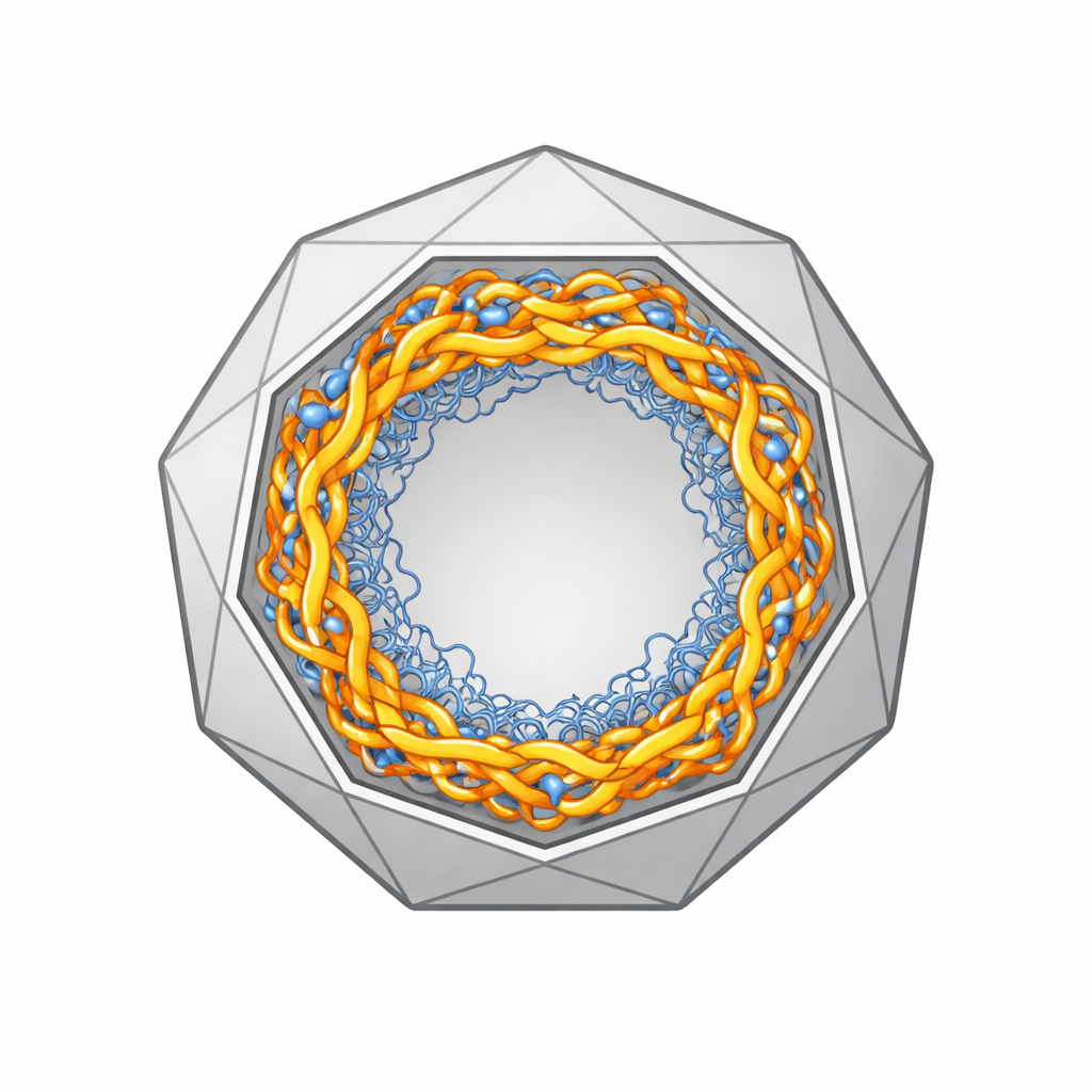
Droplets Without a Container
To understand what drives this pattern, the researchers turned to coarser, faster simulations and complementary test-tube experiments. They found that the RNA and protein tails undergo a kind of microscopic demixing known as liquid–liquid phase separation: they form dense, droplet-like condensates coexisting with more dilute surroundings. When salt levels or temperature are low, electrostatic attraction between the positively charged tails and negatively charged RNA is strong, and the condensates become more pronounced and patchy. Raising salt or temperature weakens these attractions, making the RNA layer more uniform. Similar behavior appears even when the tails are anchored to a flat surface instead of a curved capsid, and when they are mixed with short RNA fragments in bulk solution, supporting the idea that this phase-separation tendency is an intrinsic property of the mixture.
Hidden Order in a Flexible Genome
The dense patches formed by this process do more than just crowd molecules together. Within them, the RNA is more likely to fold back on itself, forming short double-stranded segments and hairpin structures, while nearby regions remain as flexible single strands. Simulations show that under conditions that promote phase separation, the number of base-paired segments rises sharply, and many of these double-stranded stretches align in parallel, forming ordered arrays. These ordered “islands” are threaded together by softer, more mobile single-stranded linkers, giving the genome a tree-like architecture that is compact yet not rigid. When the authors disrupt the charged tails, cut them loose from the shell, or neutralize their charges, both the phase separation and the base-pairing are greatly reduced. This indicates that the capsid’s inner surface, through its tethered tails, actively sculpts the genome’s higher-order shape.
Keeping the Viral Copy Machine Moving
HBV must convert its RNA genome into DNA inside the sealed capsid, a process carried out by a viral enzyme called polymerase. During this conversion, the polymerase has to jump between distant sites along the RNA, moves that depend on long-range base pairing and on the enzyme’s ability to roam through the interior. When polymerase is added to the simulations, the phase-separated, hollow-shell arrangement of RNA creates open channels in the center of the capsid where the enzyme prefers to reside and diffuse rapidly. At the same time, the organized RNA structure supports more long-distance base-pairing contacts, which are thought to guide the polymerase’s template switches. If phase separation is suppressed by neutralizing the protein tails, the RNA fills the interior more uniformly, wraps tightly around the polymerase, and slows its motion. 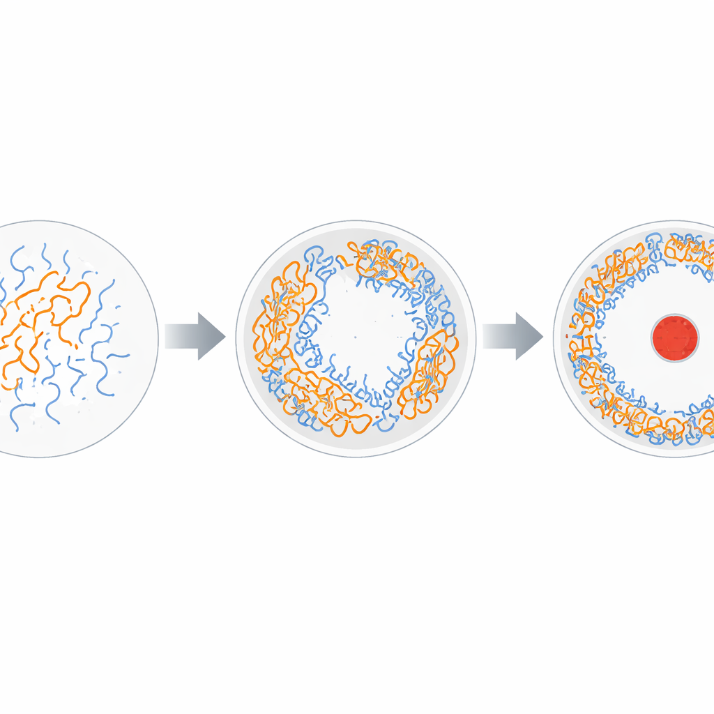
Why This Matters for Treating Hepatitis B
Taken together, these results suggest that HBV uses a fundamental physical principle—phase separation—to solve a design problem: how to fit a long genome into a tiny shell while keeping it orderly enough for accurate copying and loose enough for enzymes to move. The virus achieves this by forming a hollow layer of RNA–protein condensate along the shell wall, dotted with micro-domains of ordered hairpins and flexible linkers, and by leaving a more open interior for the polymerase. Because this organization depends sensitively on charge balance and salt conditions, it may be possible to design drugs or peptides that disrupt the condensate’s formation or stability. Targeting this physical layer of genome organization could provide a fresh route to antiviral therapies that complement approaches aimed directly at viral enzymes or entry steps.
Citation: Bian, Y., Pan, H., Mao, J. et al. Structural organization of HBV pgRNA genome driven by phase separation in capsid confinement. Nat Commun 17, 2940 (2026). https://doi.org/10.1038/s41467-026-69689-2
Keywords: hepatitis B virus, viral genome organization, liquid-liquid phase separation, RNA condensates, capsid structure