Clear Sky Science · en
Integrated histopathology of the human pancreas throughout stages of type 1 diabetes progression
Why the hidden landscape of the pancreas matters
Type 1 diabetes is usually described as the immune system attacking insulin-making cells, but what actually happens inside the human pancreas over time has been hard to see. This study uses advanced imaging and computer analysis to look across tens of thousands of tiny cell clusters in donated human pancreases, tracing how their size, makeup, and immune cell contacts change from early risk stages through long-standing disease. The work offers a more complete picture of how type 1 diabetes unfolds inside the organ, and suggests new ways to track and perhaps one day slow the process.
Looking closely at tiny cell islands
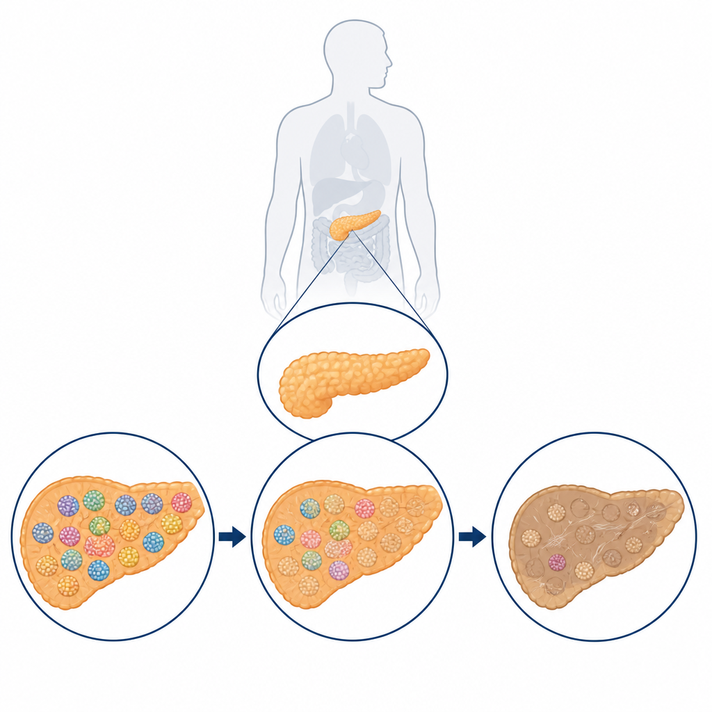
The pancreas contains thousands of small clusters called islets that house hormone-producing cells, including beta cells that make insulin and alpha cells that make glucagon. The researchers used a staining method that allows many different hormones and immune cells to be visualized one after another on the same tissue slice. They then scanned entire slices at high magnification and fed the images into open-source software that could automatically outline each islet, measure its shape and size, record which hormone-producing cells were present, and count nearby immune cells. This approach allowed them to study about 25,000 individual islets from donors without diabetes, donors who carried type 1 diabetes–related antibodies but normal blood sugar, donors at disease onset, and donors with many years of established disease.
How islets change as type 1 diabetes advances
By comparing these groups, the team found that islet health declines in several coordinated ways. As expected, donors with clinical type 1 diabetes had lost most of their beta cells and overall islet mass, but the study showed that alpha and delta cells are also reduced, while certain other hormone-producing cells remain relatively stable. Islets became more crowded with cells yet more irregular in shape, suggesting structural damage as disease progresses. A key pattern was that smaller islets, including many that normally lack alpha cells, tend to be lost early, while some larger islets temporarily retain more beta cells before succumbing later. Despite regional quirks in anatomy, such as a pancreas area rich in a hormone called pancreatic polypeptide, the overall organization of islet types and their decline looked surprisingly similar across different parts of the organ.
Early warning signs before symptoms appear
In donors who had diabetes-related antibodies but no symptoms, the pancreas already showed several warning signals. There was a clear increase in immune cells gathered around islets, and a drop in certain beta cell products such as proinsulin and a companion hormone called IAPP, even when insulin staining itself looked fairly normal. The relative number of one major group of “standard” islets shrank, replaced by somewhat larger, more stressed clusters. Using a pattern-finding tool borrowed from single-cell genomics, the team sorted islets into distinct clusters based on their structure, hormone content, and immune cell contacts. They observed that the balance among these islet clusters shifts stepwise from health, to antibody-positive risk, to early disease and then to long-duration diabetes, revealing a largely organ-wide process rather than isolated trouble spots.
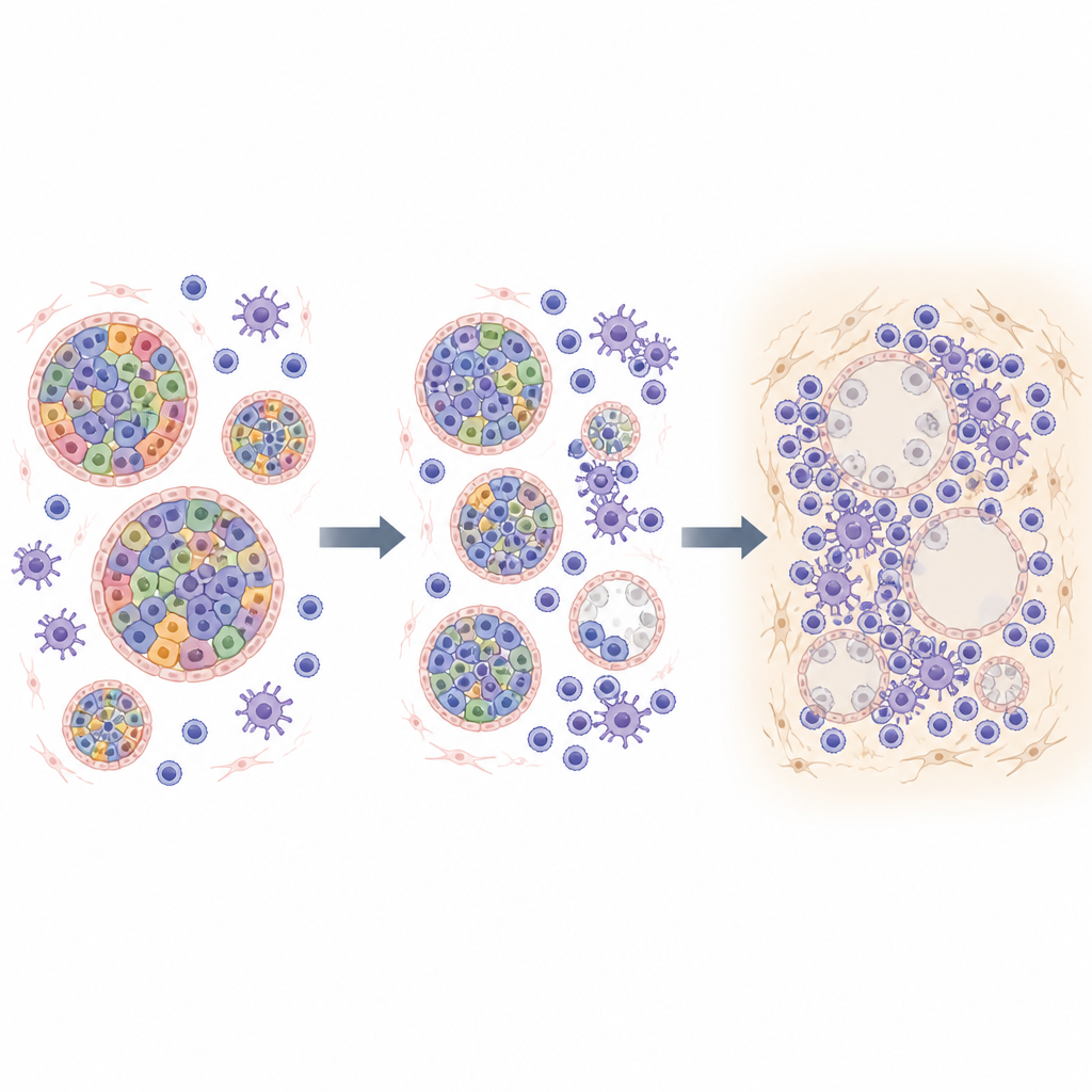
Immune attack and the wider tissue neighborhood
The classic sign of type 1 diabetes in tissue slices is insulitis, where many immune cells crowd into a few islets. This study confirms that such hot spots do occur, mainly in a particular subset of larger insulin-containing islets, but it also shows that they represent only a fraction of the immune activity. Even islets without obvious immune clusters often had reduced proinsulin and IAPP, suggesting that damage can continue after immune cells have moved on or outside of the plane captured in a single slice. By mapping the locations of islets across whole sections, the authors found that islets become more widely spaced over time, as if islands of damaged or empty islets expand and merge. Immune-rich islets tend to sit within broader neighborhoods of elevated immune presence, again pointing to a distributed, multifocal process inside the pancreas.
What this means for understanding type 1 diabetes
To a lay observer, this work reframes type 1 diabetes as a slowly shifting landscape inside the pancreas in which many islets across the organ change in concert, rather than a few being struck at random. The study suggests that subtle changes in islet makeup and rising immune presence are already widespread in people who have diabetes-linked antibodies but normal blood sugar. Over time, smaller islets disappear first, larger ones deform and lose beta cells, and the overall map of islets becomes sparser and more irregular. This integrated view of structure, cell types, immune activity, and spatial layout provides a revised natural history of type 1 diabetes that may guide future efforts to monitor risk, time interventions, and design therapies aimed at protecting as many remaining islets as possible.
Citation: van der Heide, V., McArdle, S., Nelson, M.S. et al. Integrated histopathology of the human pancreas throughout stages of type 1 diabetes progression. Nat Commun 17, 4293 (2026). https://doi.org/10.1038/s41467-026-68610-1
Keywords: type 1 diabetes, pancreatic islets, beta cells, immune cells, pancreas imaging