Clear Sky Science · en
AI-assisted analysis of early fluid dynamics following aflibercept 8 mg in treatment-naïve neovascular AMD
Why this matters for aging eyes
As people live longer, many develop age-related macular degeneration, a disease that slowly robs central vision and can make reading, driving, and recognizing faces difficult. This study looks at how a higher dose of an existing eye injection works in the very first days and weeks after treatment, using artificial intelligence to track tiny pockets of fluid in the back of the eye that are closely linked with vision quality.
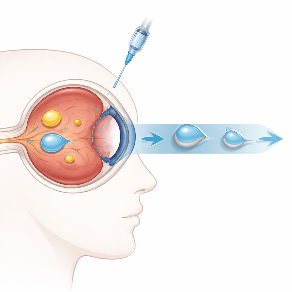
A closer look at a common cause of vision loss
The research focuses on neovascular age-related macular degeneration, a form of the disease in which fragile new blood vessels grow under the central retina and leak fluid. This leakage creates different types of fluid pockets inside and under the retina, which can blur and distort sight. Doctors already treat this condition with drugs that block a growth signal called VEGF, but patients often need frequent injections and it has been unclear exactly how quickly the higher dose of one such drug clears these fluid pockets in everyday practice.
How the study was carried out
Thirty older adults, each with one previously untreated eye affected by this condition, received three monthly injections of an 8 milligram dose of aflibercept into the eye. The team examined vision and retinal structure before treatment and at several early time points: one day, one week, two weeks, one month, two months, and three months after the first injection. They used a detailed scanning method called optical coherence tomography along with artificial intelligence software that can automatically measure the volume of three key fluid types inside a central area of the retina.
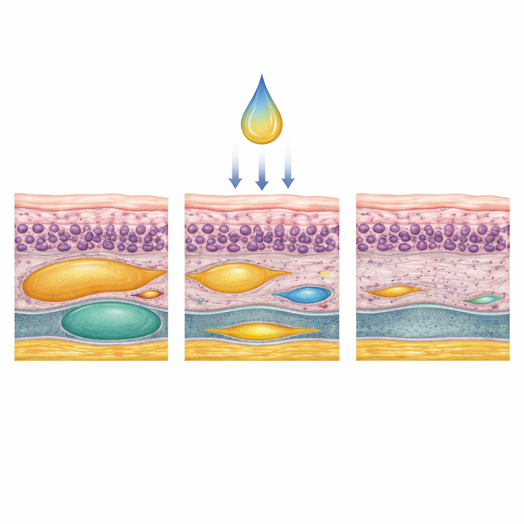
What happened to vision and retinal fluid
Vision improved quickly and stayed better over the three months. On average, patients could read about two more letters on a standard eye chart just one day after the first injection, nearly six more letters after one week, and about eleven extra letters by three months. At the same time, pockets of fluid inside the retina shrank very rapidly, dropping by about two-thirds in just one day and by more than nine-tenths within two weeks. Fluid beneath the retina also decreased strongly, though a bit more gradually, and was almost completely gone by three months in all treated eyes. Swelling in a deeper layer known as pigment epithelial detachment shrank more slowly and less completely over the same period.
Different fluid types, different healing speeds
The study found that each type of fluid followed its own timeline of improvement. Fluid trapped within the retinal tissue itself responded fastest and most completely, and the amount left after two weeks was linked to how much vision improved by three months. Fluid lying in a space just under the retina also cleared well, but at a slightly different pace depending on the pattern of abnormal vessels. The bulging deeper layer, which can be filled with either clear fluid or fibrous tissue, showed the slowest and most variable change, especially when more solid tissue was present. Overall swelling of the central retina steadily decreased, mirroring these fluid changes.
How artificial intelligence helped
Because the artificial intelligence software could measure tiny changes in fluid volume scan by scan, the researchers gained a detailed picture of how the drug acts over days rather than only months. This precise tracking suggested that early drying of fluid inside the retina may serve as a useful early sign that vision is likely to improve. The technology also allowed the team to compare responses among different patterns of diseased blood vessels and fluid types, offering clues about which patients might do well with longer gaps between injections and which might need closer follow-up.
What this means for patients and doctors
In simple terms, the higher dose of aflibercept dried out harmful retinal fluid quickly and improved sight within days in this small group of patients, with continued gains over three months. Not all fluid behaved the same way, and some deeper swelling remained, but the fast and strong response of fluid inside and just under the retina suggests that this dosing strategy could support fewer visits while still protecting central vision. By combining frequent eye scans with artificial intelligence analysis, doctors may be able in the future to fine-tune treatment plans early in the course of therapy, aiming to keep vision stable while reducing the burden of repeated injections.
Citation: Veritti, D., Sarao, V., Martin, A.A. et al. AI-assisted analysis of early fluid dynamics following aflibercept 8 mg in treatment-naïve neovascular AMD. Eye 40, 979–985 (2026). https://doi.org/10.1038/s41433-026-04319-1
Keywords: age-related macular degeneration, retinal fluid, aflibercept 8 mg, optical coherence tomography, artificial intelligence in ophthalmology