Clear Sky Science · en
Oscillatory shear stress-driven endothelial-to-mesenchymal transition: a critical mechanical signal transduction mechanism in atherosclerosis progression
Why Blood Flow Patterns Matter
Atherosclerosis—the buildup of fatty, fibrous plaques inside arteries—is the root cause of most heart attacks and strokes. Yet these dangerous plaques do not appear randomly along blood vessels: they cluster at bends, curves, and branch points. This review article explains why those spots are so vulnerable. It focuses on how a particular kind of disturbed blood flow, called oscillatory shear stress, can reprogram the cells lining our arteries, pushing them toward a more aggressive, scar-forming state that helps plaques grow and destabilize.
Quiet Stretches and Turbulent Corners
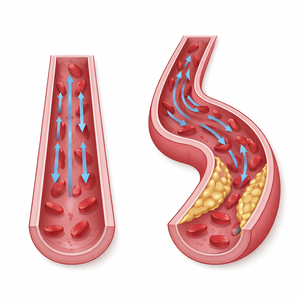
Blood vessels are constantly exposed to mechanical forces. In straight, unbranched regions, blood moves in a smooth, one-way stream that creates steady “laminar” shear stress along the vessel wall. This steady force helps keep endothelial cells—the thin layer of cells lining the artery—calm, well-organized, and protective. In contrast, at curves and branch points the flow becomes disturbed and partly reverses direction, generating oscillatory shear stress. Under these choppy conditions, endothelial cells no longer behave like a tight, uniform shield: instead, they proliferate, become inflamed, and are more likely to let fats and immune cells slip into the vessel wall, fostering the early stages of atherosclerosis.
When Lining Cells Change Identity
A central theme of the article is endothelial-to-mesenchymal transition, or EndMT. In this process, the normally flat, cobblestone-like endothelial cells gradually lose their orderly shape and specialized barrier function, then adopt features of mesenchymal cells—cells that are more spindle-shaped, mobile, and good at producing structural proteins. Studies of human atherosclerotic plaques show many cells that carry both endothelial markers and mesenchymal markers, a fingerprint of EndMT in action. The extent of this mixed identity correlates with plaque severity and instability: thin-capped, rupture-prone plaques contain more cells that have fully or partially undergone this transition.
Evidence from Animal and Cell Models
Animal experiments help connect disturbed flow to EndMT and plaque growth. In mice, researchers can alter carotid artery blood flow by placing a small cuff around the vessel or tying off several branches. These surgical tricks create regions of low and oscillatory shear stress upstream of the constriction, where the inner vessel layer thickens, plaques form quickly, and endothelial cells begin to express mesenchymal traits. In cultured human endothelial cells exposed to oscillatory shear in the lab, similar changes emerge: cells lose their tight junctions, their skeleton reorganizes, their permeability rises, and they gain enhanced movement and contractile power. Together, these changes weaken the barrier, making it easier for lipids and inflammatory cells to invade and build plaques.
How Cells Sense and Translate Mechanical Force
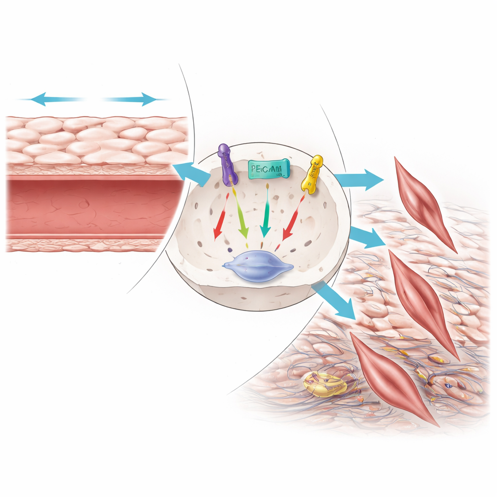
The review details the molecular “antennae” that let endothelial cells feel shear stress and convert it into biochemical responses. Ion channels such as Piezo1 and TRPV4 open in response to mechanical force, allowing calcium to surge into the cell and triggering cascades that control nitric oxide production, inflammation, and structural remodeling. Other surface proteins—including integrins, adhesion molecules like CD31, and receptors such as ALK5 and plexin D1—form complexes that sense oscillatory forces and activate pathways known to drive EndMT. A particularly important route involves TGF-β signaling, which, when over-activated by disturbed flow and epigenetic changes, turns on transcription factors like Snail and Slug that push endothelial cells toward a mesenchymal fate. The article also highlights the role of reactive oxygen species and histone modifications in amplifying these signals.
New Paths Toward Prevention and Treatment
By framing EndMT as a key link between disturbed flow and atherosclerosis, the authors argue that blocking this cellular identity switch could become a new therapeutic strategy. Experimental drugs that inhibit TGF-β-related signaling, fine-tune histone acetylation, or dampen specific mechanosensors can reduce EndMT and plaque burden in animal models. Some familiar medications, such as statins and metformin, also appear to counteract EndMT under oscillatory shear conditions. The review notes, however, that most of these approaches are still in early, preclinical stages and that EndMT is only one piece of a broader network involving lipids, inflammation, and immune cells. Still, understanding how mechanical forces reshape endothelial behavior offers a powerful lens on why plaques form where they do—and suggests that treating the “feel” of blood flow on the vessel wall may one day complement cholesterol-lowering and anti-inflammatory therapies.
What This Means for Heart Health
For a lay reader, the key message is that atherosclerosis is not just a matter of “too much cholesterol.” The physical environment inside arteries—especially how smoothly or chaotically blood flows—can reprogram the very cells that are supposed to protect us. Oscillatory shear stress at vessel bends and branches nudges these cells to behave more like scar-forming builders than like guardians of a clean, tight barrier. That shift helps plaques grow and makes them more likely to rupture, causing heart attacks and strokes. By learning how to prevent or reverse this cellular transformation, future treatments may target the disease earlier and more precisely, improving cardiovascular health beyond what is possible with current drugs alone.
Citation: Li, J., Xu, W., Ju, J. et al. Oscillatory shear stress-driven endothelial-to-mesenchymal transition: a critical mechanical signal transduction mechanism in atherosclerosis progression. Cell Death Discov. 12, 153 (2026). https://doi.org/10.1038/s41420-026-03000-6
Keywords: atherosclerosis, blood flow, endothelial cells, cellular transition, mechanotransduction