Clear Sky Science · en
Human retinal organoid model of disease-relevant photoreceptor cell death amenable to drug screening
Why protecting central vision matters
As people age, many begin to lose the sharp, central vision needed for reading, driving, and recognizing faces. One of the main culprits is dry age-related macular degeneration, a slow disease of the retina at the back of the eye. Once the light-sensing cells called photoreceptors are lost, vision cannot be restored. This study introduces a human lab-grown retina system that mimics key damage seen in the disease and can be used to search for drugs that keep these vital cells alive.
Building a tiny retina in a dish
To study how photoreceptors die in a safe and controlled way, the researchers grew miniature human retinas, called retinal organoids, from stem cells. These organoids form layered, three-dimensional tissue that closely resembles the human retina, including rods and cones that respond to light. By letting the organoids develop for about six months, the team produced tissue that behaves much like an adult retina, making it a useful stand-in for the back of the human eye when testing how injury and potential treatments affect vision cells.
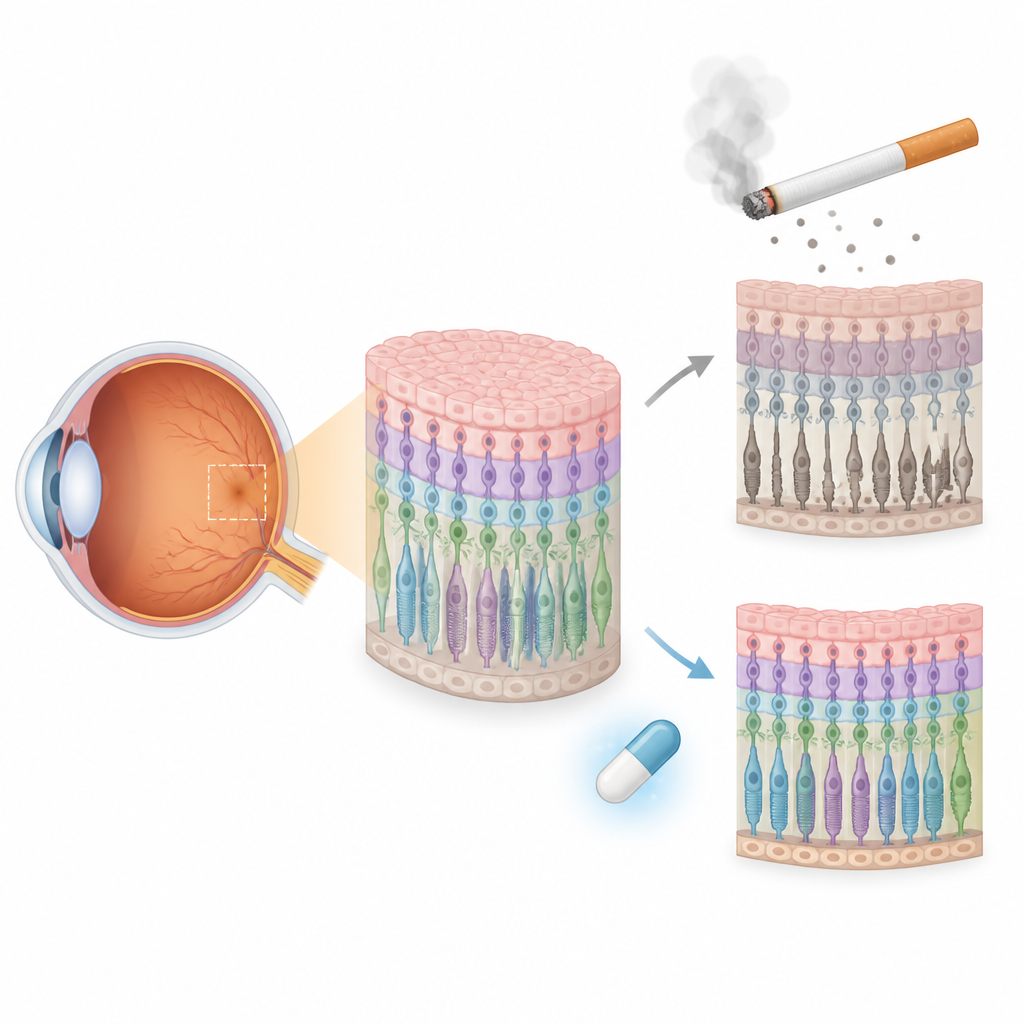
Using cigarette smoke to model eye damage
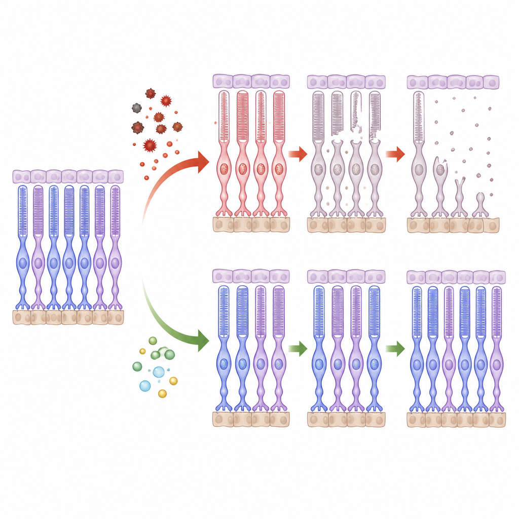
Smoking is one of the strongest lifestyle risks for macular degeneration, in part because cigarette smoke is rich in reactive chemicals that damage delicate eye tissues. The team exposed the retinal organoids to a standardized extract of cigarette smoke at different doses and times. Medium levels of extract caused clear stress and cell death without completely destroying tissue structure, closely mirroring the gradual harm seen in disease. The dying cells were located mostly in the outer nuclear layer, where photoreceptors live, showing that the model mainly targets the same cell type that fails in patients.
How stressed cells lose their power
The study dug into what happens inside photoreceptors as they are harmed. The smoke extract sharply increased the production of reactive oxygen species, unstable molecules that attack cell components. At the same time, the tiny power plants inside cells, the mitochondria, lost their normal electrical charge, a sign that they were failing. Signals linked to a self-destruct program known as the intrinsic apoptosis pathway were switched on: key executioner proteins were activated, and the balance between survival and death signals tipped toward cell loss. These changes match patterns seen in human macular degeneration and animal studies.
Iron, rusty fats, and a second type of cell death
Beyond classic cell suicide, the researchers found evidence for another, more recently described death route called ferroptosis. In the organoids, iron built up inside cells and fats in their membranes became oxidized, making the membranes unstable. The cell’s main antioxidant system, based on a molecule called glutathione, responded by ramping up production, but the ratio of its protective form dropped, revealing that defenses were being overwhelmed. Large-scale protein analysis confirmed disruptions in iron handling, redox balance, energy use, and cell waste processing, pointing to a tangled network of stress pathways similar to those implicated in macular degeneration.
Turning the model into a drug discovery tool
To make the organoid system useful for testing new treatments, the team paired it with fast, non-destructive fluorescent readouts that can be measured in living tissue. These readouts track cell death, oxidative stress, mitochondrial health, and lipid damage in many organoids at once, using plate readers suited for high-throughput screening. The investigators also showed that simpler chemical stressors such as hydrogen peroxide and sodium iodate can produce related patterns of damage, offering alternative setups when a more narrowly focused injury is desired.
What this means for future eye therapies
In simple terms, this work shows that lab-grown human retinas can be pushed into disease-like states that closely resemble what happens in dry macular degeneration, especially in the vulnerable photoreceptors. Because the system captures both classic cell suicide and iron-driven membrane damage, and can be read rapidly and repeatedly, it provides a powerful platform to test drugs that might keep light-sensing cells alive. Used alongside existing models, this human-centered approach could speed the search for treatments that not only slow tissue changes but also help preserve the sight people rely on every day.
Citation: Parween, S., Saviola, A.J., Howell, A.C. et al. Human retinal organoid model of disease-relevant photoreceptor cell death amenable to drug screening. Cell Death Dis 17, 474 (2026). https://doi.org/10.1038/s41419-026-08724-y
Keywords: age-related macular degeneration, retinal organoids, photoreceptor cell death, oxidative stress, ferroptosis