Clear Sky Science · en
Stiff matrix-induced KRTAP2-3 expression suppresses ciliogenesis via actin tension-driven chromatin remodeling
How Cells Feel Their Surroundings
Our bodies are built from tissues that can be squishy like brain or firm like bone, and living cells are surprisingly sensitive to this stiffness. This study reveals how that physical “feel” of the surroundings can switch off tiny antenna-like structures on cells called primary cilia, which help cells sense signals crucial for normal growth, development, and health.
Little Antennas On Every Cell
Primary cilia are slim, hairlike projections that stick out from many cells and act as miniature antennae. They pick up chemical and physical cues and help control processes such as fat storage, cell division, daily body rhythms, and embryo development. When these cilia are missing or faulty, organs across the body can be affected, leading to a group of disorders known as ciliopathies.
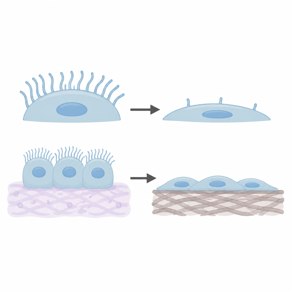
Stiff Surroundings Cut Down Cilia
The researchers grew human and mouse cells on laboratory-made gels that ranged from very soft, like brain tissue, to very stiff, like bone. They then counted how many cells carried a primary cilium. As the surface became stiffer, the fraction of ciliated cells dropped sharply, even though the length of each cilium stayed about the same. Gene activity measurements showed that groups of genes linked to cilia building became less active on stiff surfaces, while genes linked to cell scaffolding and attachment to the surface became more active.
Cell Skeleton Tension As The Middleman
To understand how stiffness sends its message inside, the team focused on actin, a key part of the cell’s internal skeleton. On stiffer surfaces, actin fibers became longer, more numerous, and more aligned, forming taut cables across the cell. When drugs were used to loosen or break up these actin fibers, the differences in cilia numbers between soft and stiff surfaces largely disappeared, and more cells regrew cilia. This showed that tension within the actin network acts as a main go-between that translates outside stiffness into cilia loss.
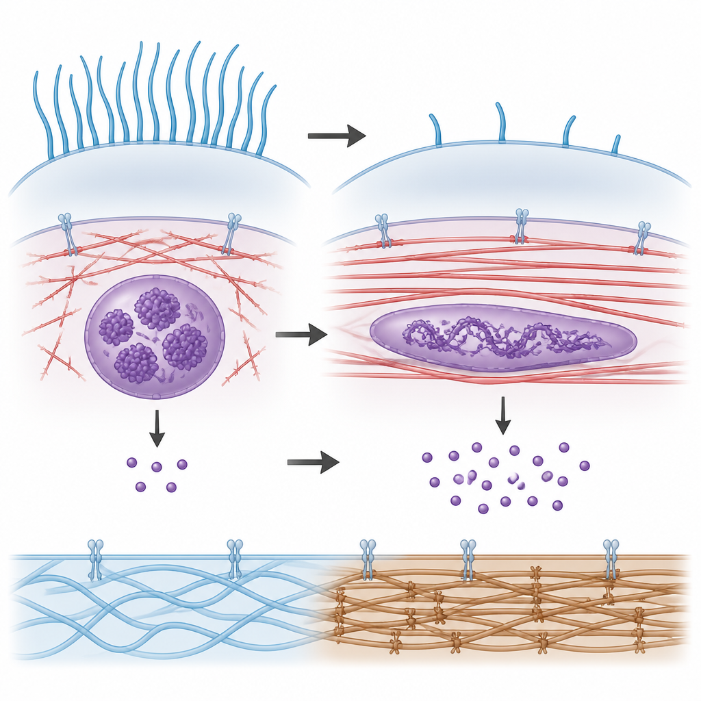
A Switch Gene That Listens To Tension
By scanning all active genes, the scientists found one, called KRTAP2-3, that was strongly turned up in cells on stiff surfaces. When they reduced KRTAP2-3 levels, cells regained their cilia even on stiff or medium surfaces. When they boosted KRTAP2-3, cells lost cilia even on soft, normally friendly surfaces. Importantly, breaking up actin fibers lowered KRTAP2-3 activity, tying this gene directly to the state of the cell skeleton. This suggests that KRTAP2-3 works as a stiffness-sensitive switch that decides whether cilia are built or suppressed.
How Shape Changes Reach The Cell’s Library
The team then asked how actin tension could change KRTAP2-3 so strongly. Using computer models and microscopy, they showed that as surfaces stiffen and actin fibers pull harder, the cell nucleus flattens and spreads. This reshaping alters how tightly packed the DNA is in certain regions. A technique that detects open stretches of DNA revealed that the region near the KRTAP2-3 gene became more accessible in cells on stiff surfaces, and this opening depended on intact actin fibers. In other words, physical pulling on the nucleus helps “open a page” in the genetic library where KRTAP2-3 sits, making it easier to read and copy.
Why This Matters For Health And Disease
Tissues naturally change stiffness during development and in diseases such as scarring and cancer. This work outlines a full chain of events: a stiff environment tightens the cell skeleton, deforms the nucleus, opens the DNA near KRTAP2-3, increases this gene’s activity, and in turn suppresses the formation of primary cilia. Understanding this physical-to-genetic pathway may help explain why cilia are often lost in stiff, diseased tissues and could one day guide ways to restore cilia-related signaling by tuning the cell’s mechanical environment or its internal tension.
Citation: Chen, X., Yi, L., Xie, G. et al. Stiff matrix-induced KRTAP2-3 expression suppresses ciliogenesis via actin tension-driven chromatin remodeling. Cell Death Dis 17, 443 (2026). https://doi.org/10.1038/s41419-026-08678-1
Keywords: primary cilia, matrix stiffness, mechanotransduction, actin cytoskeleton, chromatin remodeling