Clear Sky Science · en
Changes in the cortical GABAergic inhibitory system in a Spinal Muscular Atrophy mouse model
Why brain balance matters in a muscle disease
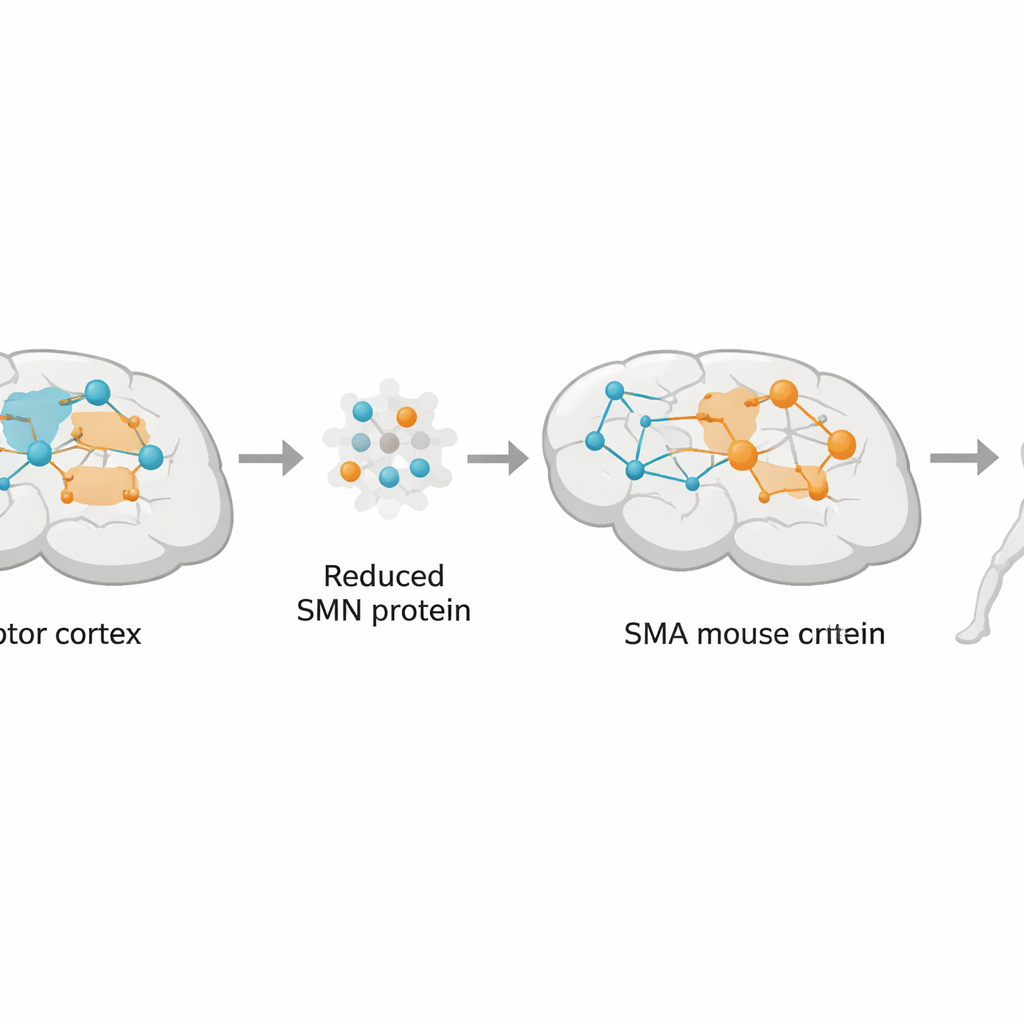
Spinal muscular atrophy (SMA) is best known as a devastating childhood disease that weakens the muscles and can be fatal. For years, scientists focused mainly on the spinal motor neurons that directly control movement. This study asks a different question: what if part of the problem also lies higher up, in the brain’s own movement center? By looking at how “brake” cells in the motor cortex malfunction in a severe SMA mouse model, the researchers uncover a hidden layer of disease that may help explain symptoms and point to new treatment strategies.
A fragile truce between brain signals
Normal movement depends on a truce between two kinds of nerve-cell activity in the cortex: excitatory signals that drive neurons to fire and inhibitory signals that rein them in. Many brain disorders, from epilepsy to Parkinson’s disease, are now understood as disruptions of this excitatory–inhibitory balance. In SMA, patients and animal models show structural and functional changes in the motor cortex, suggesting that the “brake system” may be altered there as well. Yet most research has concentrated on the spinal cord. The authors set out to test whether inhibitory signaling in the sensorimotor cortex is disturbed in SMA, and whether this disturbance is linked to the loss of a protein called SMN, whose deficiency causes the disease.
The brain’s brake cells under stress
Using a combination of brain imaging, molecular assays, and electrical recordings, the team examined the sensorimotor cortex of SMA mice at different disease stages. They focused on GABA, the main inhibitory messenger in the brain, and on a key class of GABA-producing cells known as parvalbumin interneurons, which act as fast, precise brakes on motor output. In late-stage SMA mice, the density of GABA-positive neurons and the intensity of GABA signal were reduced, especially in the deep layer 5 of the cortex where output neurons that command the spinal cord reside. The enzymes that make GABA (GAD65 and GAD67) were also decreased, and parvalbumin interneurons showed fewer branches and smaller cell bodies, hinting at a loss of inhibitory strength exactly where it matters most for controlling movement.
Weaker synapses and scrambled chemistry
To see how these structural changes affect function, the researchers measured inhibitory electrical currents received by layer 5 pyramidal neurons. In SMA mice, these cells experienced fewer action-potential-driven inhibitory events, even though the size of each event was larger, a pattern consistent with a failing but partially compensating brake system. Microscopic analysis confirmed fewer inhibitory synaptic contacts on these neurons, both in brain tissue and in simplified cell cultures. At the same time, chemical profiling of the cortex revealed a late-stage buildup of glutamine, a precursor used by neurons to make both glutamate and GABA. Rather than a simple shortage of GABA in the tissue as a whole, these findings point to a misrouting of the glutamine–glutamate–GABA cycle within local circuits.
Astrocytes, transporters, and the SMN connection
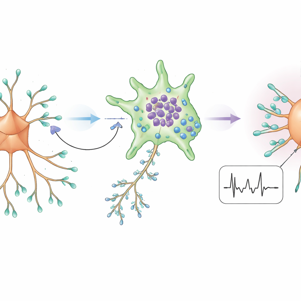
Because the SMN protein helps regulate RNA processing and thus protein production, the team asked how its loss might distort this chemical cycle. They found that a glutamine transporter mainly made by astrocytes—a support cell type that shuttles fuel to neurons—was reduced in the SMA cortex. Another transporter that pulls GABA back into astrocytes was increased, and astrocytes in SMA cultures accumulated more GABA-like signal than neighboring neurons. When SMN levels were experimentally lowered in otherwise normal neurons, their GABA signal dropped; when SMN was boosted with the SMA drug Nusinersen in culture, glutamine transport and GABA levels improved. Together, these results suggest that SMN deficiency disrupts neuron–astrocyte cooperation, starving inhibitory neurons of the raw materials they need while encouraging astrocytes to sequester more of the available GABA.
What this means for people with SMA
To a lay reader, the message is that SMA is not just a disease of the spinal cord but also of brain circuits that shape movement. The study shows that in a severe SMA mouse model, the motor cortex gradually loses part of its braking system: specialized inhibitory cells shrink, make less GABA, form fewer synapses, and are hampered by faulty support from nearby astrocytes. This leaves output neurons more vulnerable and may worsen motor symptoms even as current SMN-boosting therapies prolong survival. The work helps explain why drugs that enhance inhibitory signaling have sometimes shown benefits in SMA and suggests that future treatments may need to combine SMN restoration with strategies that directly stabilize cortical inhibition and neuron–astrocyte metabolism.
Citation: Menduti, G., Ferrini, F., Caretto, A. et al. Changes in the cortical GABAergic inhibitory system in a Spinal Muscular Atrophy mouse model. Cell Death Dis 17, 285 (2026). https://doi.org/10.1038/s41419-026-08520-8
Keywords: spinal muscular atrophy, motor cortex, GABA inhibition, interneurons, neuron–astrocyte metabolism