Clear Sky Science · en
Decoding cellular communication networks and signaling pathways in bone, skeletal muscle, and bone-muscle crosstalk through spatial transcriptomics in a young male mouse
How Bones and Muscles Talk to Each Other
Our bones and muscles do far more than hold us up and move us around. They constantly exchange chemical messages that help set our strength, metabolism, and ability to recover from injury. This study used a powerful mapping technique to see, for the first time in high detail, how different cells in bone and nearby skeletal muscle communicate in place inside a young mouse leg. Understanding this hidden conversation could eventually shed light on conditions like osteoporosis, muscle wasting, and age-related frailty.
Looking Inside Living Tissues in Place
Instead of grinding tissue into a mush of cells, the researchers kept a thin slice of mouse femur and its attached leg muscle almost exactly as it sits in the body. They then applied spatial transcriptomics, a method that measures which genes are switched on while preserving where each signal came from. Using a commercial platform, they captured thousands of tiny spots across the section, each recording the activity of hundreds of genes. By aligning these molecular readouts with standard microscope images, they could tell whether a spot came from solid bone, the spongy inner bone, bone marrow, or muscle.
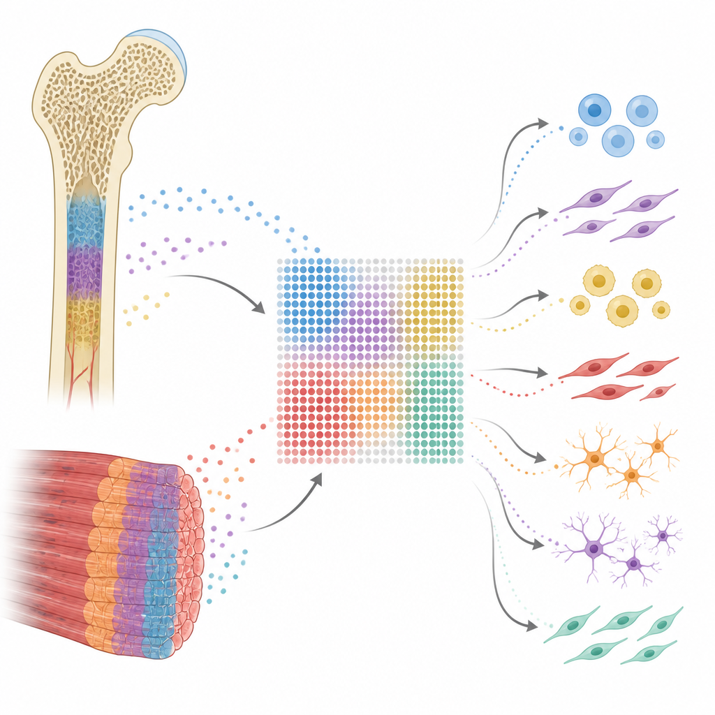
Who Lives Where in Bone and Muscle
Because each spot can hold several cells, the team used computational tools to estimate which cell types were present and in what proportions. They identified eight main players, including red blood cell precursors, blood vessel cells, bone-forming osteoblasts, muscle fibers, immune cells such as monocytes and macrophages, stem-like support cells, and fat cells. As expected, osteoblasts and stem cells clustered along the hard and spongy bone surfaces, blood-forming and vessel cells filled the marrow, and muscle fibers dominated the muscle region. This produced a detailed “cell atlas” of the bone–muscle unit, confirming that spatial transcriptomics can resolve complex architecture in such dense tissues.
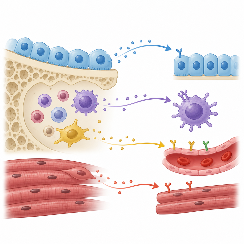
Tracing the Web of Cellular Messages
Next, the researchers focused on how these cells might talk to each other. They examined pairs of genes that form classic signaling units: one cell makes a secreted or surface “ligand” protein, and another cell displays the matching receptor. With a specialized analysis tool, they inferred which cell types were most active as senders and receivers of these messages. Blood-forming and vessel cells, along with monocytes and macrophages, sat in the middle of dense communication networks. Osteoblasts sent and received many signals and often talked to themselves in feedback loops. Muscle fibers showed moderate but clear connections with bone and immune cells, hinting that under calm, healthy conditions their crosstalk is present but not extreme.
Key Pathways that Link Bone, Muscle, and Blood
The team highlighted several families of molecules that seemed especially important. Collagen-based signals, which help build and organize the tissue scaffold, flowed strongly to and from osteoblasts and shaped the interfaces between bone, marrow, and muscle. Another protein, osteopontin, linked bone cells with blood and immune cells and is known to affect bone renewal and muscle repair. Monocytes and macrophages relied on thrombospondin and fibronectin pathways to influence osteoblasts, blood vessels, and muscle fibers, underscoring their role as coordinators of tissue remodeling. In muscle, message routes involving tenascin and VEGF stood out, connecting muscle fibers to blood vessels and immune cells in ways that support blood supply and healing.
Checking the Map Against Reality
To make sure the predicted conversations were not just statistical artifacts, the scientists used multiplex immunostaining, a method that labels specific proteins in tissue sections with glowing tags. They confirmed that several key ligand and receptor partners, such as certain collagen and tenascin proteins and their binding partners, appeared together in the right cell types at bone–muscle boundaries. They also turned to independent single-cell datasets from mouse and human bones. Even though those datasets lacked muscle, most of the same signaling pathways and many of the same ligand–receptor pairs reappeared, suggesting that the communication map is robust and shared across species.
What This Means for Bone and Muscle Health
This work offers an initial, spatially resolved blueprint of how bone, muscle, blood-forming cells, and immune cells coordinate their activities in a healthy young mouse. It shows that bone-forming cells, immune cells, and muscle fibers use overlapping sets of scaffold proteins and growth factors to keep tissues strong, supplied with blood, and ready to repair damage. While the study does not yet address disease directly, it lays the groundwork for future research into how these same signaling routes change with aging, injury, or metabolic disorders, and how tuning them might one day help preserve both bone and muscle function.
Citation: Qiu, C., Li, Y., Gong, Y. et al. Decoding cellular communication networks and signaling pathways in bone, skeletal muscle, and bone-muscle crosstalk through spatial transcriptomics in a young male mouse. Bone Res 14, 55 (2026). https://doi.org/10.1038/s41413-026-00520-w
Keywords: bone muscle crosstalk, spatial transcriptomics, cell communication, ligand receptor signaling, musculoskeletal biology