Clear Sky Science · en
Abnormal signal transmission in white matter revealed by resting-state communication connectivity in Alzheimer’s disease: A comprehensive cross-sectional and longitudinal study
Why this study matters to families and caregivers
Alzheimer’s disease slowly robs people of memory and independence, but by the time symptoms are obvious, much damage has already occurred in the brain. This study looks for earlier warning signs hidden in how brain areas "talk" to each other through their wiring—the white matter pathways. By tracking subtle changes in these communication routes over time, the research suggests new ways to detect Alzheimer’s earlier and monitor whether treatments are working.
Looking at the brain’s wiring, not just its surface
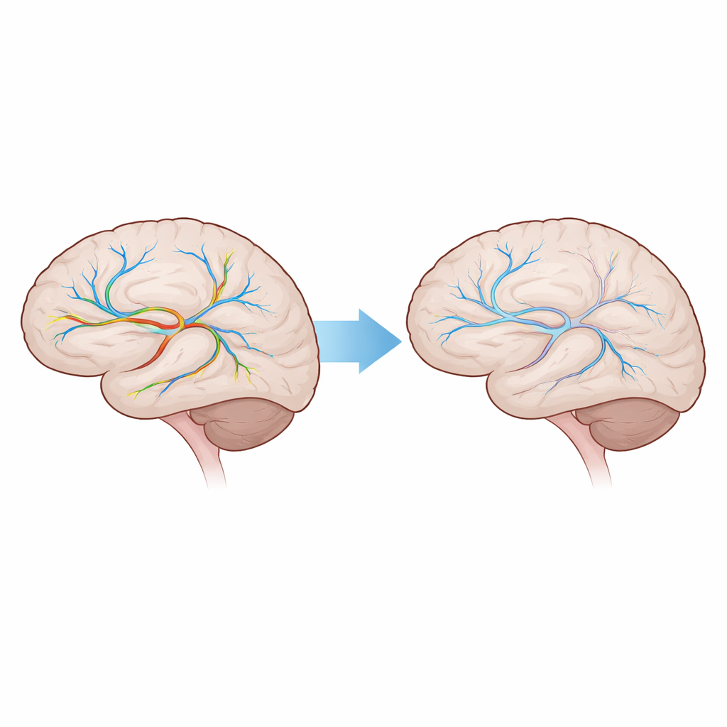
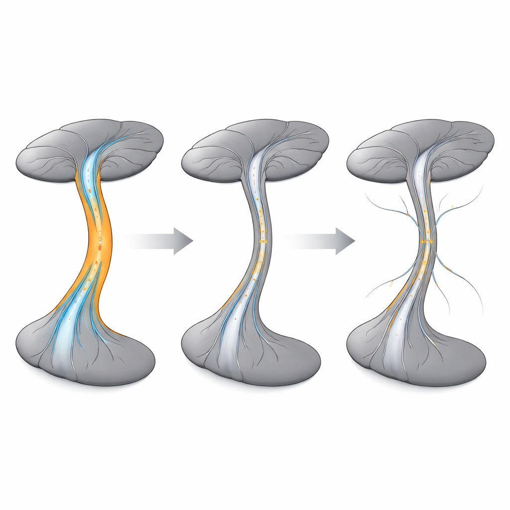
Most brain scans used in Alzheimer’s research focus on gray matter, the outer layer where nerve cells process information. Traditional "functional connectivity" methods look at how the activity of two gray matter regions rises and falls together. But signals do not jump directly between these regions—they travel along white matter bundles, the brain’s internal wiring. Until recently, standard methods largely ignored what happens inside these pathways. The authors set out to fill this gap by asking: how does Alzheimer’s disease alter the actual routes that messages take through white matter?
A new way to measure brain conversations
The team used resting-state functional MRI, a scan that measures natural fluctuations in blood oxygen levels while a person lies still in the scanner. They applied a newly developed measure called "communication connectivity," which looks at activity in three places at once: one gray matter region, a white matter bundle, and a second gray matter region. Instead of simply pairing two surface areas, this approach evaluates how well a specific white matter pathway supports the conversation between them. For each of 82 gray matter regions and 48 major white matter bundles, the researchers built detailed maps showing how strongly different parts of the brain communicate through each pathway.
Following changes across the Alzheimer’s spectrum
The study drew on data from 169 older adults in the Alzheimer’s Disease Neuroimaging Initiative, including people who were cognitively normal, those with early and late mild cognitive impairment, and those with Alzheimer’s dementia. All had at least two brain scans taken 9 to 30 months apart. The researchers compared the communication patterns between groups at the first scan and then tracked how each person’s patterns changed over time. While traditional gray matter–to–gray matter connectivity looked surprisingly similar across groups, the new communication maps revealed clear shifts as the disease progressed, especially in certain deep white matter tracts.
Key signal routes falter early and keep worsening
Among many bundles examined, two stood out: the right retrolenticular part of the internal capsule and the right posterior corona radiata—deep pathways that help link processing areas across the brain. In people with early mild cognitive impairment, communication through these routes was already measurably weaker than in healthy older adults. The decline continued in late mild cognitive impairment and was most pronounced in those with Alzheimer’s dementia. Importantly, this pattern appeared both when comparing different people at one point in time and when following the same individuals over time, suggesting a consistent signature of disease progression rather than a one-time fluctuation.
What this could mean for early diagnosis and treatment
For non-specialists, the main message is that Alzheimer’s does not only shrink brain tissue; it also disrupts the hidden highways that carry signals between regions. By focusing on how information moves along specific white matter bundles, this study identifies early breakdowns in key routes that could serve as sensitive markers of the disease. If confirmed in larger and longer studies, these communication measures could help doctors detect Alzheimer’s earlier, track how quickly it is advancing, and evaluate whether new therapies are truly protecting the brain’s wiring. In short, listening more carefully to how the brain’s internal networks communicate may offer a clearer window into the disease’s earliest and most treatable stages.
Citation: Guo, Y., Huang, W., Xiong, X. et al. Abnormal signal transmission in white matter revealed by resting-state communication connectivity in Alzheimer’s disease: A comprehensive cross-sectional and longitudinal study. Transl Psychiatry 16, 120 (2026). https://doi.org/10.1038/s41398-026-03883-0
Keywords: Alzheimer’s disease, white matter, brain connectivity, resting-state fMRI, early biomarkers