Clear Sky Science · en
Integrative transcriptomics and electrophysiological profiling of hiPSC-derived neurons identifies novel druggable pathways in Koolen-de Vries Syndrome
Why this brain disorder study matters
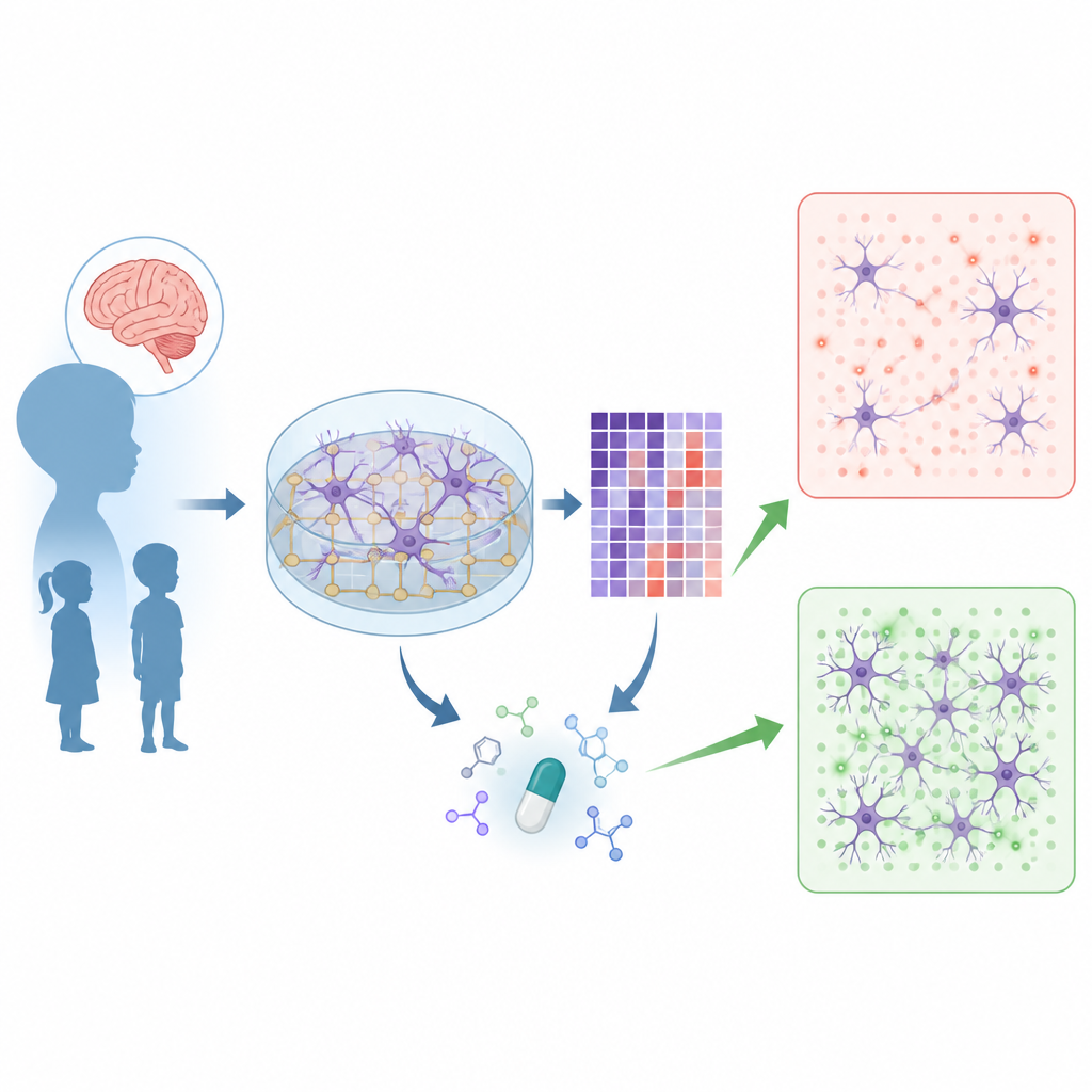
Koolen-de Vries Syndrome is a rare genetic condition that causes developmental delay, learning difficulties, and low muscle tone. Families currently have no specific medical treatment, only supportive care. This study shows how scientists can grow nerve cells from patients in the lab, watch how their electrical activity differs from typical cells, and use those differences to hunt for medicines that might restore more normal brain cell communication.
Growing patient brain cells in a dish
Researchers began by turning skin cells from people with Koolen-de Vries Syndrome and from unaffected volunteers into induced pluripotent stem cells, which can be coaxed to become many cell types. They then pushed these stem cells to develop into excitatory brain cells and grew them on special plates containing tiny electrodes that record electrical signals. Over several weeks, these lab-made networks started to fire in coordinated bursts, mimicking the way groups of neurons talk to each other in the brain.
Finding what goes wrong in cell conversations
When the team compared patient-derived networks with those from controls, they found that Koolen-de Vries neurons fired less often in synchronized bursts and did so in a more irregular, jittery pattern. At the same time, these neurons formed fewer synapses, the contact points where cells exchange signals. To understand why, the scientists created an approach they call MEA-seq, in which they record network activity and then immediately measure which genes are turned on or off in the very same cultures. By matching electrical features with gene activity levels, they could pinpoint molecules that appear to shape how well the network fires.
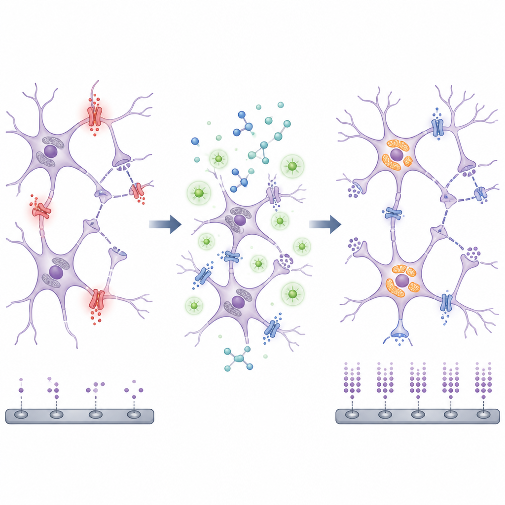
A chloride channel and tired energy factories
One standout finding was a gene called CLCN4, which makes a chloride channel protein. Higher levels of this gene in patient neurons were linked to weaker, less frequent bursts and longer pauses between them. When the researchers dialed down CLCN4 in patient cells, the timing and strength of network bursts shifted back toward control-like patterns, and synapse numbers improved. The study also uncovered a strong link between genes involved in mitochondria, the cell’s energy factories, and healthy firing patterns. Follow-up experiments showed that patient neurons had poorer mitochondrial respiration and reduced ability to make cellular fuel, along with lower reliance on sugar breakdown, suggesting an overall energy shortfall.
Screening existing drugs with gene signatures
Armed with the gene activity patterns from patient neurons, the team turned to a large public database that catalogues how thousands of drugs change gene expression in human cells. They asked which compounds tend to reverse the Koolen-de Vries–like gene signature, especially the mitochondrial and network-related changes. From this computational screen they selected ten existing or experimental medicines with likely effects on energy metabolism or related pathways and tested them for weeks on patient-derived neuron networks to see whether electrical activity became more regular and synchronized.
Promising effects of a natural antioxidant
Two compounds, fasudil and phloretin, stood out for their ability to nudge abnormal network activity closer to control levels. Phloretin, a plant-derived antioxidant found in apples, produced the most consistent benefits across several patient cell lines. It increased the fraction of spikes that occurred within organized bursts, boosted burst rates in some lines, and reduced the variability between bursts. Gene analyses showed that both drugs enhanced programs linked to neuronal projections, the long processes that host synapses, and phloretin also boosted pathways related to energy metabolism. In parallel, phloretin restored synapse density to near-control levels and lowered markers of oxidative stress in patient neurons.
What this could mean for future treatment
This study does not yet offer a therapy for people with Koolen-de Vries Syndrome, but it charts a concrete path forward. By combining electrical recordings and gene readouts from patient-derived neurons, the researchers could trace how a missing copy of the KANSL1 gene leads to disturbed network rhythms through altered ion channels, fewer synapses, and underpowered mitochondria. Using the same data, they identified existing compounds, such as phloretin, that partly normalize these problems in the dish. In the long run, this integrated strategy may speed the development and testing of targeted treatments not only for Koolen-de Vries Syndrome but also for other neurodevelopmental conditions where brain cell networks fall out of sync.
Citation: Verboven, A.H.A., Puvogel, S., Latour, B.L. et al. Integrative transcriptomics and electrophysiological profiling of hiPSC-derived neurons identifies novel druggable pathways in Koolen-de Vries Syndrome. Mol Psychiatry 31, 3558–3575 (2026). https://doi.org/10.1038/s41380-026-03482-x
Keywords: Koolen-de Vries Syndrome, hiPSC neurons, neuronal networks, mitochondrial dysfunction, drug repurposing