Clear Sky Science · en
Pipeline integrating cultural heritage X-ray computed radiography and computed tomography data using DICOM
Why Old Objects Need New Kinds of X‑Rays
Museums and archives increasingly rely on X‑ray and CT scanners to peek inside fragile treasures without touching them. But while the images may look like hospital scans, the data needs are very different: curators must track an object’s history, ownership, and ethics alongside the pixels, and they need files that will still be trustworthy decades from now and easily shared on the web. This paper introduces a new digital pipeline, called DICOCH, that reshapes a medical imaging standard so it can safely carry the stories and legal conditions of cultural heritage objects into the future.
The Problem with Scanning the Past
Conservators already use non‑destructive X‑ray methods—computed radiography (CR) and computed tomography (CT)—to study how artworks and artifacts are built, damaged, or repaired. Yet the resulting data are often scattered: images in one folder, spreadsheets in another, and separate PDF reports. That fragmentation makes it easy to lose links between an object and its context, such as when and where it was made, who owns it, and what may or may not be shown online. Existing imaging standards from medicine and industry, while powerful, do not fully capture this cultural information or complex rights, and they rarely connect cleanly to modern web viewers.
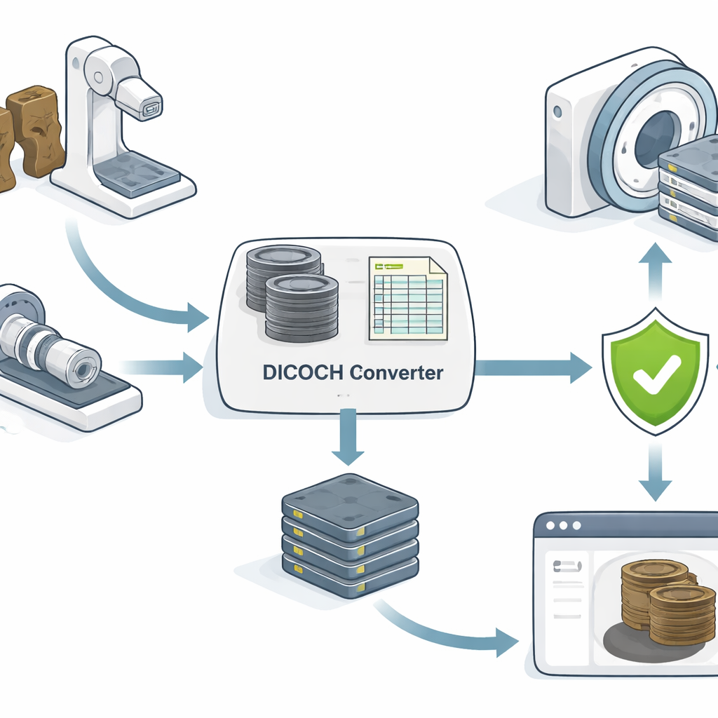
Building a Single Trustworthy Container
The author proposes DICOCH (“DICOM for Cultural Heritage”), a three‑stage workflow named Generation–Validation–Publication. It starts by converting raw X‑ray or CT images plus tabular metadata into standardized DICOM files, the same kind of container used in hospitals. A carefully designed “private” section inside each file is reserved for heritage‑specific details like provenance, material type, conservation history, copyright holder, and usage license. The system also records how raw gray values from industrial scanners are linearly mapped into the familiar medical CT scale, while keeping the original numbers untouched. In the next stage, an official validation tool checks every file against the DICOM rulebook, enforcing a zero‑error policy. Finally, the pipeline automatically creates web‑ready image derivatives and IIIF manifests so that the same source files can feed both professional medical viewers and open cultural heritage viewers in a browser.
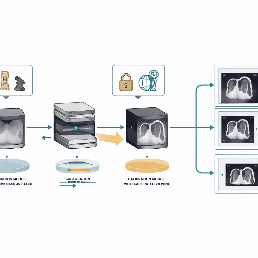
Trying It on a Historic Wooden Mask
To test the idea, the study uses CT and X‑ray scans of the Andong Hahoe Mask, a 12th‑century Korean wooden mask designated a national treasure. The pipeline converts very large 2D radiography images and 3D CT slices into DICOM while writing the mask’s official status, material description, and management numbers directly into the embedded heritage metadata block. For the CT data, DICOCH encodes the slice geometry so that standard radiology software can reconstruct and navigate through the volume. It then applies a simple, well‑known linear transform so that air, wood, paint, and metal fasteners appear with consistent contrast on medical displays, but always links these “nominal” values back to the original raw data and to a dedicated calibration record that can be refined later.
Proving It Works Across Systems
When the DICOCH files were opened in a commercial medical viewer, they passed all checks: spatial measurements, scrolling through slices, pixel readouts, and contrast adjustments all worked as expected, and the custom heritage information remained attached. In contrast, vendor‑supplied files from the same machines showed multiple standard violations, from missing required fields to conflicting descriptions, which sometimes caused glitches or mis‑scaled images. The same DICOCH files also drove web viewers through IIIF manifests, allowing deep zoom and side‑by‑side comparison in a browser while still exposing structured heritage and rights data pulled from the embedded private group.
What This Means for Digital Heritage
For non‑specialists, the key result is that DICOCH turns what used to be a jumble of images and documents into a single, self‑describing package that computers and people can both trust. It keeps the most accurate version of the scan intact, clearly separates it from how the image is displayed, and locks in the object’s story, ownership, and viewing rules so they cannot silently drift away from the pixels. Because the tools and tag dictionaries are openly shared, other institutions can reuse and extend this approach, potentially forming the basis of a future official standard. In practical terms, that means a CT scan made today of a fragile artifact can be reliably re‑analyzed in a hospital‑grade environment and viewed around the world online, without sacrificing either scientific rigor or cultural responsibility.
Citation: SONG, JI. Pipeline integrating cultural heritage X-ray computed radiography and computed tomography data using DICOM. npj Herit. Sci. 14, 211 (2026). https://doi.org/10.1038/s40494-026-02480-0
Keywords: cultural heritage imaging, X-ray computed tomography, digital preservation, DICOM standard, IIIF web access