Clear Sky Science · en
Noncanonical function of epigenetic reader YTHDF1 inhibits MASLD progression by maintaining peroxisomes and mitochondrial homeostasis
Why this liver story matters
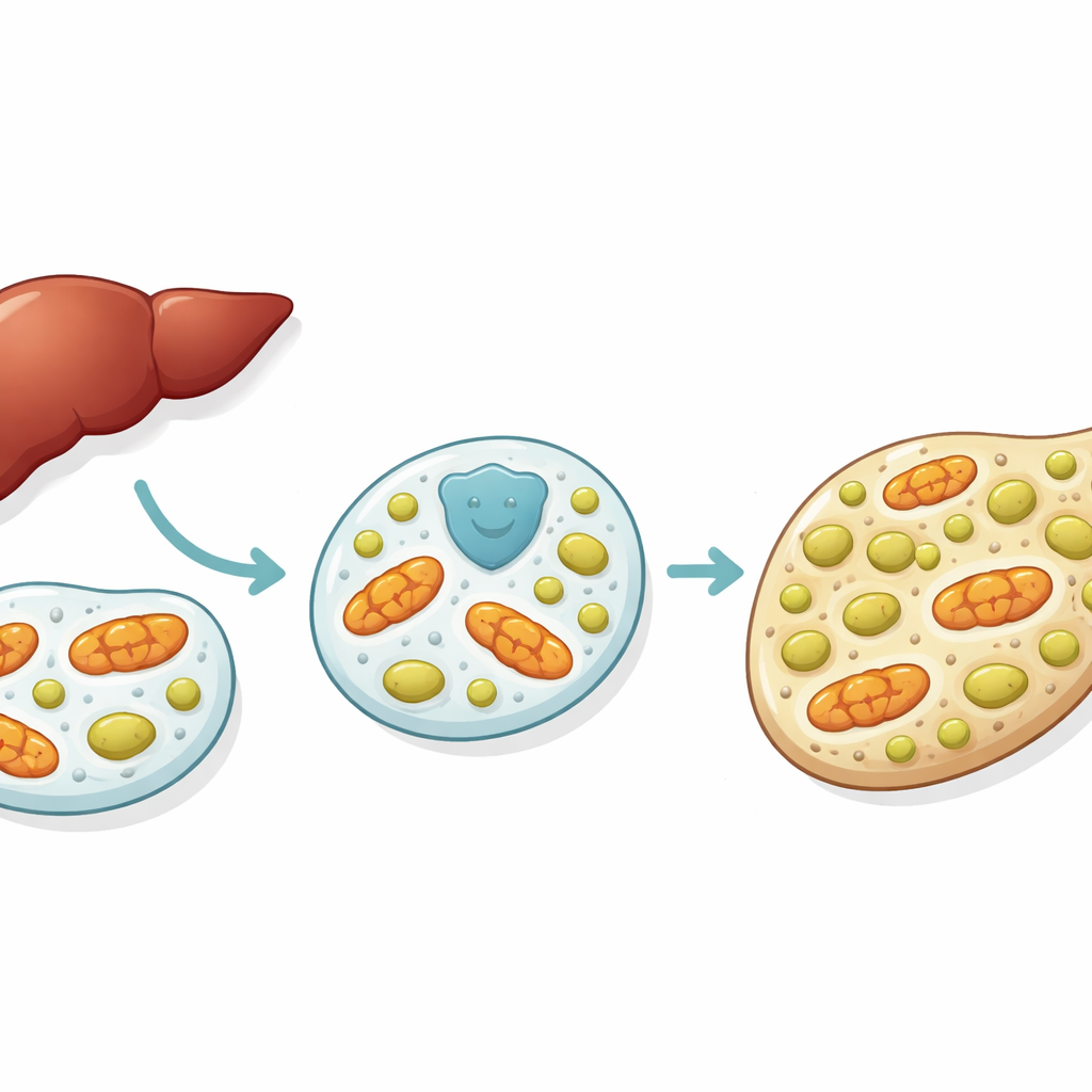
Many people live with extra fat in their liver without realizing it. This condition, now called metabolic dysfunction-associated steatotic liver disease, is tightly linked to obesity and type 2 diabetes and can silently progress to cirrhosis and liver cancer. The study behind this article uncovers how a little-known cellular helper protein, YTHDF1, helps protect liver cells from fat overload by keeping two vital structures—peroxisomes and mitochondria—in balance. Understanding this hidden defense system could point the way toward new strategies to slow or stop fatty liver disease.
A growing problem inside the liver
Fatty liver disease develops when the liver takes in more fat than it can safely handle. Extra fat and sugars from an unhealthy diet push liver cells to burn more fat, which creates harmful byproducts called reactive oxygen species. These byproducts damage cell structures and trigger inflammation. Over time, the liver becomes swollen, scarred and less able to do its many jobs. Because there are currently no approved drugs that directly treat this condition, scientists are keen to find natural protective systems within liver cells that could be boosted or mimicked.
A guardian protein steps into the spotlight
The researchers focused on YTHDF1, a protein best known for how it reads chemical marks on RNA molecules and helps control protein production. They examined liver samples from people and mice with diet-induced fatty liver disease and found that YTHDF1 protein levels rose during early stages of the disease even though its RNA levels stayed the same. In mice specially engineered to lack YTHDF1 only in liver cells, a high-fat diet led to larger, fattier livers, more liver damage markers in the blood and more inflammation than in normal mice. When YTHDF1 was added back to these livers using a viral delivery method, fat build-up and tissue injury were partly reversed, suggesting that YTHDF1 acts as a local guardian against diet-driven damage.
Keeping tiny cleaning stations under control
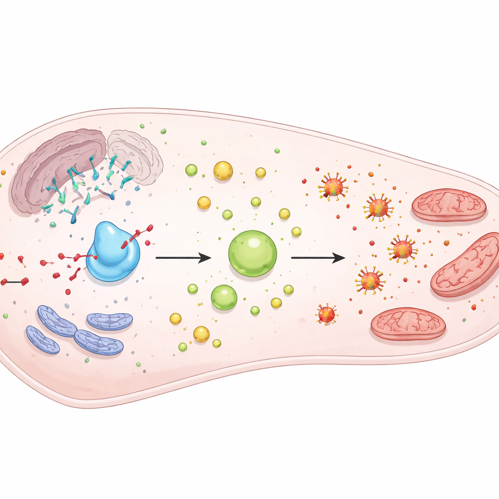
One of YTHDF1’s key protective roles involves peroxisomes, tiny compartments that help break down fatty acids. Using broad surveys of RNA and protein levels, the team found that when YTHDF1 was missing, many peroxisome-related proteins rose without changes in their RNA, hinting at an unusual control step. In particular, levels of ACOX1, a rate-setting enzyme for fat burning inside peroxisomes, increased strongly under high-fat conditions. This led to extra production of reactive oxygen species and overactivation of a growth pathway that suppresses “lipophagy,” the cell’s process for clearing fat droplets. The researchers showed that YTHDF1 helps form stress granules—temporary droplets inside cells that store certain RNAs during stress—and that ACOX1 RNA becomes trapped in these droplets. With less YTHDF1, fewer stress granules form, more ACOX1 is made and peroxisomes become overactive, which paradoxically worsens fat accumulation.
Protecting the cell’s power stations
The study also revealed that YTHDF1 physically resides within mitochondria, the cell’s energy factories, and binds to several mitochondrial proteins. Loss of YTHDF1 in liver and liver-like cells led to swollen, fragmented mitochondria with disrupted inner folds, reduced oxygen use and lower energy output, especially under fat overload. One important partner of YTHDF1 is SLC25A11, a transporter that moves the antioxidant glutathione into mitochondria. Without YTHDF1, SLC25A11 levels dropped, total glutathione handling changed and harmful mitochondrial reactive oxygen species climbed, triggering a wave of stressed, partly completed “self-eating” structures around damaged mitochondria. However, the final fusion of these structures with cellular recycling centers was impaired, so damaged mitochondria piled up instead of being cleared.
How the liver’s shield is tuned
Finally, the team asked how the cell controls YTHDF1 itself. They discovered that the protein carries a small chemical tag—methylation—on a particular lysine building block. This tag makes YTHDF1 less stable and more prone to breakdown. Under early high-fat stress, methylation on this site fell while YTHDF1 protein levels rose, hinting that the cell may temporarily stabilize YTHDF1 as a defensive move. In advanced disease, YTHDF1 levels declined, which may reflect later shifts in methylation or other controls, and could help explain why protection eventually fails.
What this means for people with fatty liver
Put simply, YTHDF1 helps liver cells cope with fat overload by locking away a powerful fat-burning enzyme when it would cause harm and by keeping the cell’s power stations healthy and well supplied with antioxidant defenses. When YTHDF1 is lost or its balance is disturbed, peroxisomes and mitochondria go out of tune, leading to more oxidative stress, blocked fat clearing and faster progression of fatty liver disease. While this work was done in cells and mice, it highlights YTHDF1 and its partners as potential targets for future treatments aimed at restoring the liver’s own protective systems rather than simply blocking fat entry.
Citation: Mu, C., Tan, J., Wang, Y. et al. Noncanonical function of epigenetic reader YTHDF1 inhibits MASLD progression by maintaining peroxisomes and mitochondrial homeostasis. Exp Mol Med 58, 1172–1186 (2026). https://doi.org/10.1038/s12276-026-01686-3
Keywords: fatty liver disease, MASLD, mitochondria, peroxisomes, YTHDF1