Clear Sky Science · en
Reversibility and therapeutic feasibility of DNM1L-associated neurodevelopmental disorders
Why this rare brain disorder matters
Some children develop normally for a short time and then begin to lose skills, suffer relentless seizures, and show shrinking of the brain on scans. For families and doctors, one painful question is whether the damage is already fixed at birth or whether there is still a chance to intervene. This study looks at a rare genetic condition tied to a gene called DNM1L and asks: are the changes in brain cells permanent, or can they be reversed if we act early enough?
When a cell’s power factories go wrong
DNM1L carries the instructions for making DRP1, a protein that helps mitochondria and peroxisomes—tiny structures that manage energy and clean-up inside cells—divide and keep their shape. In children with DNM1L-associated encephalopathy, this system goes awry, leading to highly fused, elongated organelles instead of a balanced network. The result is a broad set of problems, including developmental delay, seizures that resist medication, and visible brain shrinkage, especially of the outer brain and the bridge between the two hemispheres. Because current treatments only ease symptoms, understanding when and how this damage unfolds is crucial for designing therapies that do more than just manage seizures.
Clues from patients and engineered mice
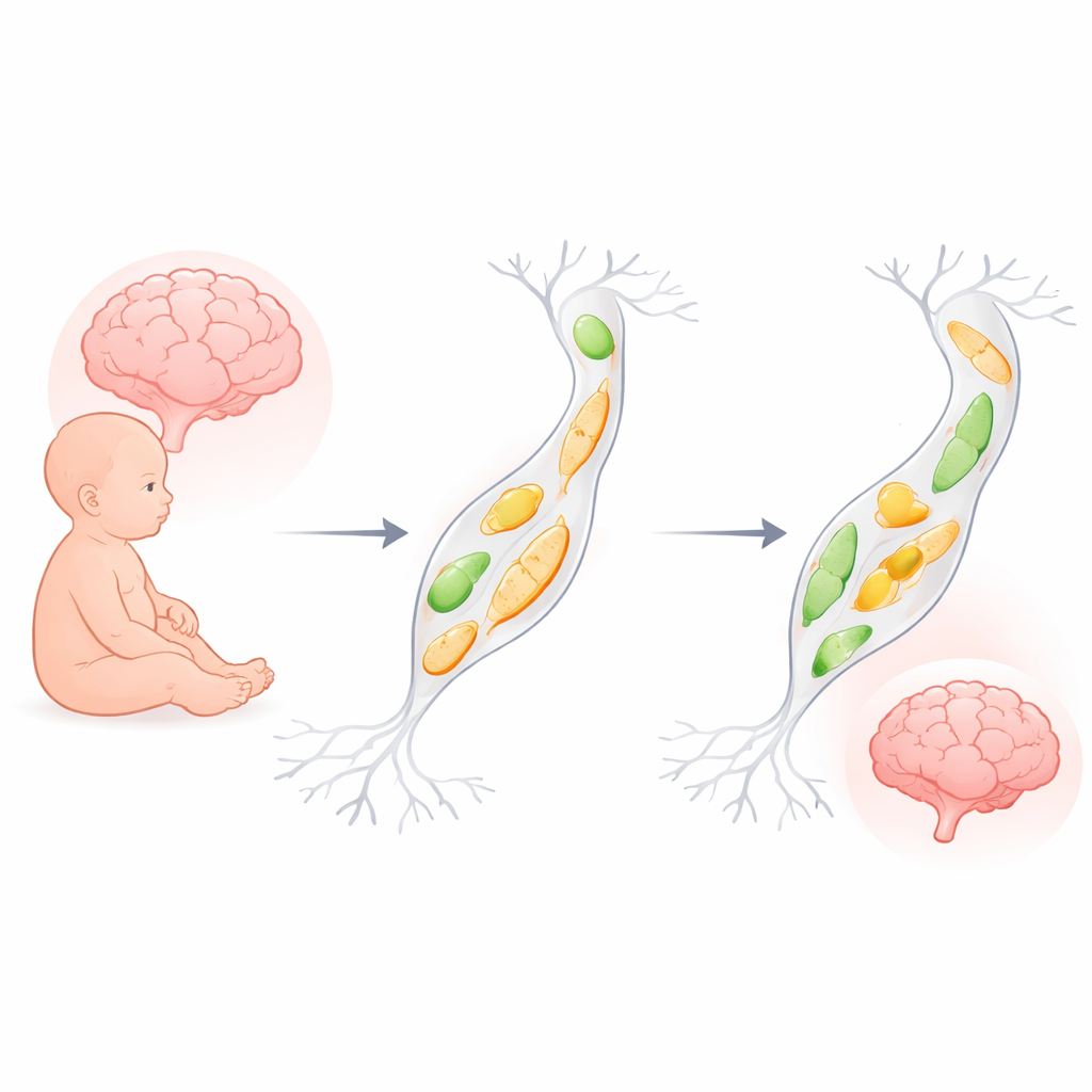
The researchers first identified two children with new, harmful changes in DNM1L that affect a stalk-like region of the DRP1 protein. Computer modeling suggested that these mutations destabilize the protein’s normal ring-like structure, and experiments in human neural precursor cells confirmed that the altered DRP1 causes the overly fused mitochondria and elongated peroxisomes seen in other DNM1L disorders. To see how this plays out in a living brain, the team introduced human DNM1L variants into a subset of developing mouse brain cells before birth. As the mice matured, those neurons showed reduced survival, simplified wiring across the corpus callosum, and thinning of this key communication highway—changes that mirror the brain scans of affected children.
Damage that increases after birth, not before
One striking finding was timing. During fetal stages, the mutant gene did not strongly alter how many brain cells were being born or how quickly they divided. Instead, major problems appeared after birth: during the first postnatal week, neurons carrying the mutant DNM1L were much more likely to die, while neighboring glial cells were spared. This selective loss of neurons offers a cellular explanation for the progressive brain shrinkage seen in patients. It also highlights a vulnerable window just after birth when neurons are integrating into circuits and may be more likely to be removed if they function abnormally.
Testing reversibility in human stem-cell–derived neurons
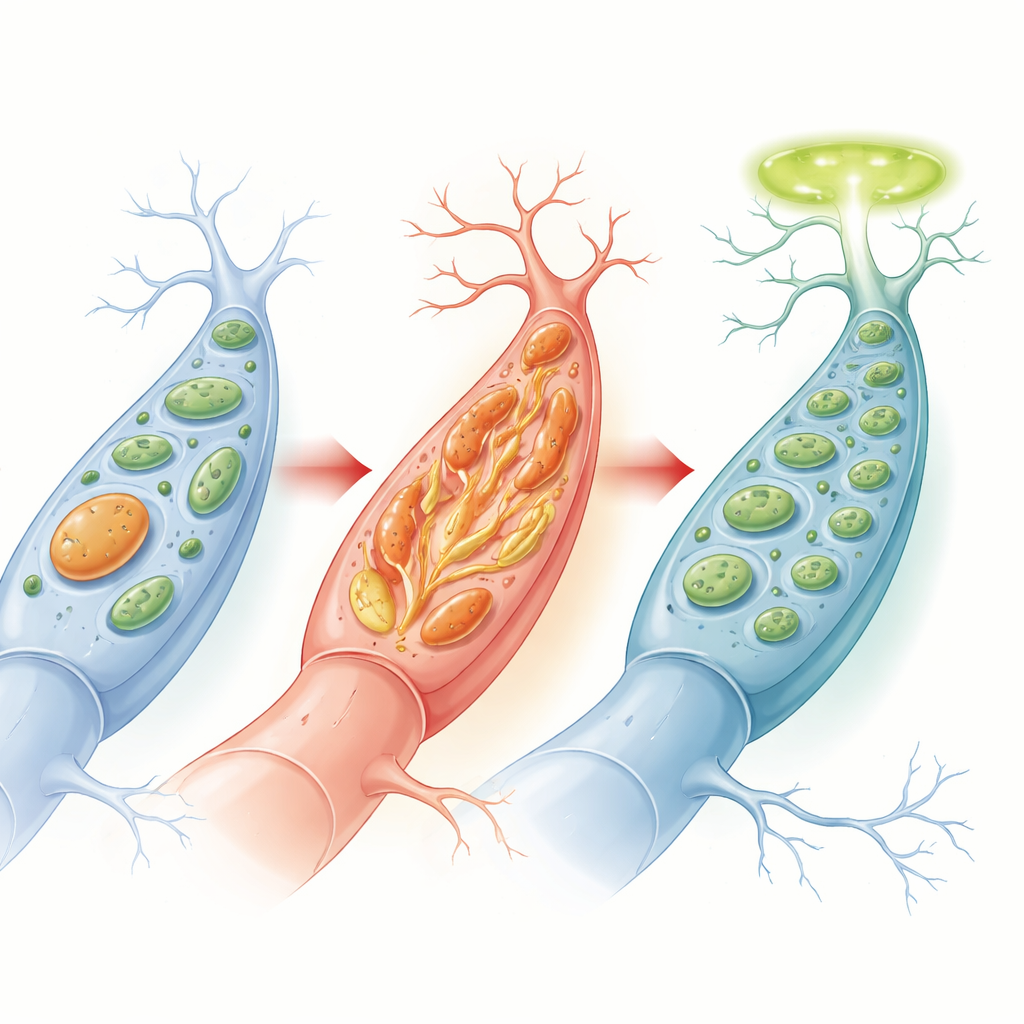
To probe whether these changes could be undone, the team built a human stem cell model in which DNM1L could be switched off and then back on using a drug. Turning DNM1L off in neural precursor cells and their descendant neurons triggered wide-ranging shifts in gene activity: stress and cell-death programs were turned up, while genes needed for synapses and electrical signaling were turned down. Yet when DNM1L expression was restored—either while cells were still precursors or even after they had become mature neurons—more than three quarters of these gene-expression changes moved back toward normal. Pathways tied to cell death, stress, and key electrical channels were especially reversible, suggesting that many harmful signals are not locked in and that neurons retain a surprising degree of molecular flexibility.
Boosting cell power to protect newborn neurons
Armed with this insight, the researchers looked for biological pathways that were both strongly disturbed by DNM1L loss and largely reversible. Mitochondrial biogenesis—the process by which cells build new mitochondria—stood out. Enhancing a master regulator of this pathway, PGC1α, in the mouse brain countered neuron loss driven by several DNM1L mutants. A drug called bezafibrate, known to activate the same pathway, also protected vulnerable mouse neurons grown in dishes and, importantly, improved survival of affected neurons in newborn mice when given during the first postnatal week. These results do not prove that bezafibrate itself will help children, but they show that strengthening the cell’s energy-making capacity can blunt the core cellular damage.
What this means for future treatments
For families facing DNM1L-related disorders, this work offers cautious but genuine hope. It shows that in models of the disease, most of the harmful molecular changes in neurons can be reversed if the underlying defect is corrected, even after the cells have matured. It also identifies a practical treatment angle—boosting mitochondrial biogenesis—that can partially protect neurons during a critical early-life window. While more advanced animal models and clinical studies are needed to test whether such strategies can improve seizures or development, the message is clear: in at least some genetic brain disorders, early postnatal life may represent a real therapeutic window, not a point of no return.
Citation: So, K.H., Kim, S.H., Jang, S. et al. Reversibility and therapeutic feasibility of DNM1L-associated neurodevelopmental disorders. Exp Mol Med 58, 755–767 (2026). https://doi.org/10.1038/s12276-026-01660-z
Keywords: DNM1L, mitochondrial dynamics, neurodevelopmental disorder, neuronal loss, mitochondrial biogenesis