Clear Sky Science · en
Non-invasive jaundice detection using spectral-band expansion from RGB images and direct hyperspectral images
Why yellow eyes matter
Most people think of jaundice simply as yellow skin or eyes, but behind that color shift lies a buildup of a blood pigment called bilirubin that can signal serious liver or blood problems. Today, checking bilirubin usually means drawing blood in a clinic or hospital, which can be painful, slow, and hard to access for newborns, older adults, and people in remote areas. This study asks a deceptively simple question with far‑reaching consequences: can an ordinary phone camera, helped by smarter image analysis and a lab‑grade optical camera, spot jaundice reliably enough to guide care without a needle?
Looking for clues in the white of the eye
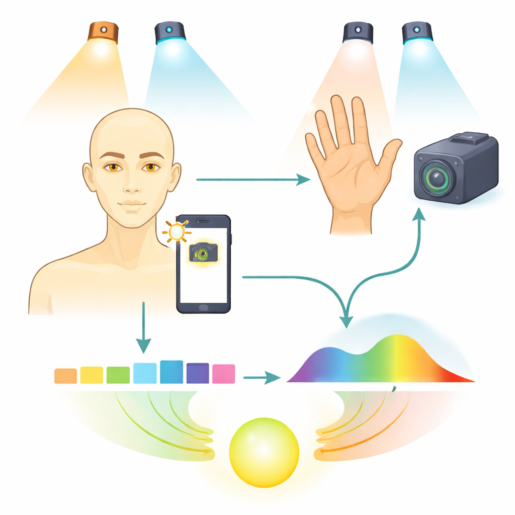
The team focused first on the sclera—the white part of the eye—because its color is less affected by sun exposure and skin tone than the skin itself. They collected close‑up eye photos from 47 patients under two common kinds of indoor lighting: warm halogen lamps and cooler fluorescent tubes. To make sure that differences in room light did not masquerade as disease, each image was passed through a two‑step “normalization” process that anchors colors to bright and dark reference spots in the same picture. The researchers then expanded each ordinary red‑green‑blue (RGB) image into 13 carefully chosen color bands that capture subtle shifts between blues, greens, yellows, and oranges—precisely the region where jaundice shows up to the human eye.
Teaching a phone to estimate blood chemistry
From each eye image, the 13‑band color fingerprint of the sclera was fed into a compact machine‑learning model called JaundiceAI‑Mobile. Rather than trying to guess a simple yes/no answer, the system learned to predict the same numerical jaundice index that doctors obtain from blood tests. Training used 90 images with known blood results, and the model was tuned separately for the two lighting types. Under fluorescent‑style lighting, which resembles many office and home environments, the predictions matched the laboratory measurements extremely closely: the statistical fit (R²) was 0.988, and the linear correlation was 0.9945, meaning the phone‑based estimates almost perfectly tracked rising and falling bilirubin levels across the study group.
Seeing beyond human vision with hyperspectral images
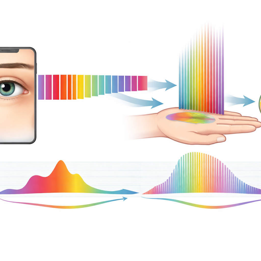
While phones see only three broad color channels, a specialized hyperspectral camera can record dozens of narrow wavelength bands from each pixel, including invisible near‑infrared light. The researchers used such a camera to examine patients’ palms, extracting tiny patches of smooth skin, mixed skin, and skin creases. By converting raw interferogram videos into full spectra, they obtained 141 wavelength points per spot from 400 to 1,000 nanometers. When they averaged these spectra across groups with different jaundice severities, a consistent picture emerged. In people with jaundice, skin reflected less blue‑green light (below about 550 nanometers) but more yellow‑orange light (around 560–590 nanometers)—changes that match the classic yellow appearance. More intriguingly, in the near‑infrared range the team found new crossover points where jaundiced and healthy skin swapped which was brighter, especially around 750–850 nanometers and near 850, 950, and 980 nanometers.
Hands, creases, and hidden signals
The palm creases turned out to be particularly revealing. These folds are rich in connective tissues that can accumulate bilirubin and are less influenced by blood flow and pigment. Hyperspectral scans of the creases showed that, in visible light, jaundiced palms tended to be darker than normal ones. Yet in a narrow near‑infrared window roughly between 690 and 855 nanometers, the trend reversed and jaundiced creases reflected more light. This pattern, together with the consistent crossing points seen in both eye‑based color data and palm‑based hyperspectral data, suggests that the body’s yellowing follows a robust optical signature that can be tracked across tissues and cameras. By mapping the 13 phone‑friendly color channels onto matching hyperspectral wavelengths, the authors outline a path for “super‑resolution” models that let smartphones approximate the richer spectral view without expensive hardware.
From lab concept to everyday checkup
For patients and families, the take‑home message is that a simple photo of the eye, when processed carefully, can come surprisingly close to replacing a blood draw for gauging how jaundiced someone is—at least within the limits of this early trial. The study also shows that there is more diagnostic information in our skin than the naked eye can see, especially in near‑infrared light. Together, the high accuracy of the phone‑based predictions and the detailed hyperspectral fingerprints point toward a future in which people could monitor jaundice at home or in low‑resource clinics using familiar devices, with advanced optics and algorithms quietly translating subtle color shifts into meaningful medical insight.
Citation: Liao, WC., Lin, J.J.Y., Lu, YC. et al. Non-invasive jaundice detection using spectral-band expansion from RGB images and direct hyperspectral images. npj Biosensing 3, 22 (2026). https://doi.org/10.1038/s44328-026-00087-w
Keywords: jaundice, smartphone imaging, hyperspectral imaging, noninvasive diagnostics, bilirubin