Clear Sky Science · en
Multi-contrast laser endoscopy for in vivo gastrointestinal imaging
Seeing Hidden Trouble in the Gut
Colonoscopy and other “scope” procedures are meant to spot early signs of cancer and other diseases in the digestive tract before they become dangerous. Yet many small or subtle growths still slip past even expert eyes, in part because they barely stand out in standard camera views. This paper introduces a new kind of endoscope lighting, called Multi-contrast Laser Endoscopy (MLE), designed to make suspicious tissue pop out more clearly by revealing not just color, but also blood flow and surface shape in real time.
Why Regular Scopes Can Miss What Matters
Today’s hospital endoscopes shine bright white light and record color video with a high-definition camera. Doctors look for differences in color, texture, and shape of the thin lining (mucosa) that coats the esophagus, stomach, and colon. The problem is that early tumors and precancerous polyps can look almost identical to healthy tissue; their colors are only slightly different and their shapes only gently raised or flattened. Even a widely used “enhanced” option called narrow band imaging, which emphasizes blood vessels by using specific blue-green colors, has not meaningfully reduced the rate at which colon polyps are missed.
A New Way to Light Up the Inside
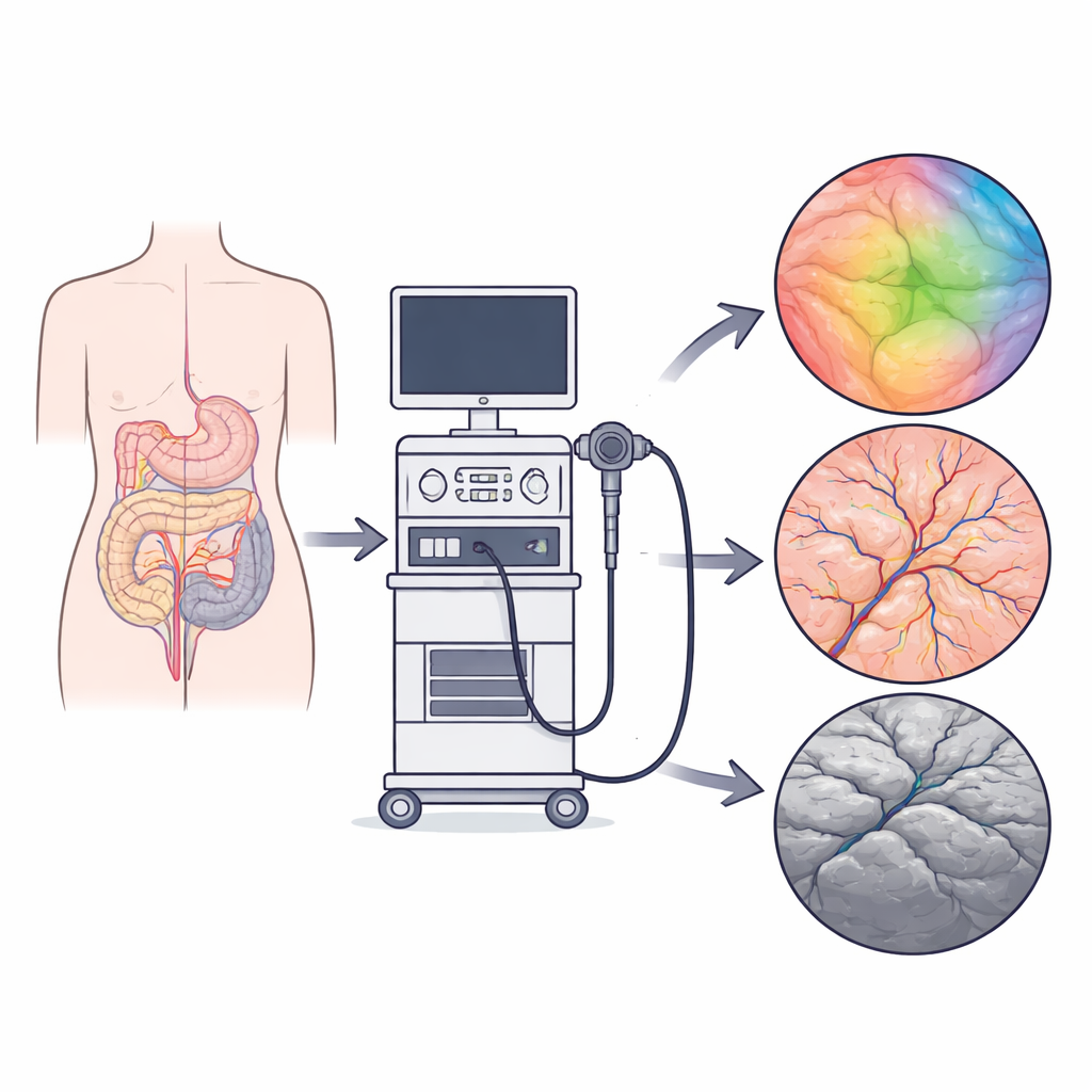
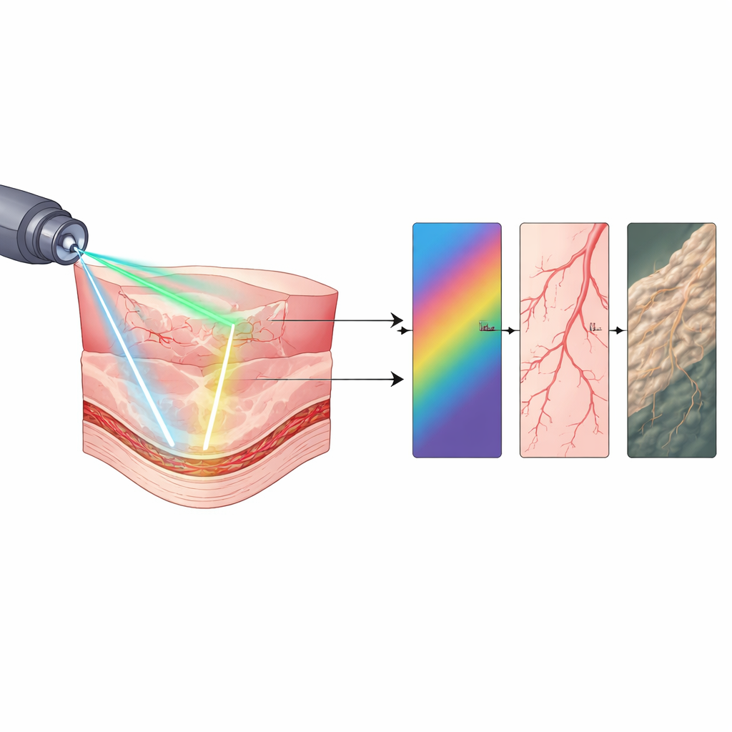
The researchers developed MLE by modifying a standard clinical colonoscope so it could accept light from a custom laser box without giving up any of its normal features. Instead of sending just broad white light, the box can rapidly switch among several carefully chosen laser colors, change how “speckled” the light is, and direct light from different angles. Inside the scope, tiny optical fibers mix this laser light with the usual hospital light, so the same camera and optics capture both. The system can toggle between ordinary images for the doctor and experimental modes for research in less than a second, all while maintaining a wide view, sharp focus over typical working distances, and full video frame rates.
Turning Light Into Extra Clues
With this flexible lighting, MLE can collect three new kinds of information during a routine procedure. First, by cycling through multiple laser colors and measuring how the tissue reflects each one, the system can map the presence of key light-absorbing molecules such as blood pigments and estimate local oxygen levels. Second, by briefly using a highly coherent laser that creates a grainy pattern called speckle, and then analyzing how that pattern blurs over time, MLE can highlight regions where blood is moving and even gauge relative flow speeds. Third, by shining light from different directions in quick sequence, MLE can reconstruct the fine hills and valleys of the mucosal surface, enhancing subtle bumps and edges that are hard to see under flat, uniform lighting.
From Bench Tests to Real Patients
The team first checked that these new modes were accurate and reliable. Color charts showed that the spectral measurements matched a laboratory spectrometer, and a simple arm experiment confirmed that the oxygen maps changed as expected when blood flow was briefly cut off and then restored. Tiny channels flowing with bead-filled liquid mimicked blood vessels and demonstrated that speckle analysis could pick up changes in flow, while silicone models of the colon confirmed that directional lighting could faithfully recover surface height and small topographic features. The researchers then brought MLE into real colonoscopy exams for 20 patients, capturing images from 31 confirmed precancerous polyps while doctors performed standard care. Compared with normal white light and narrow band imaging, the MLE-based color rendering increased color differences between polyp and surrounding tissue by about five-fold, and the surface-shape maps roughly doubled the contrast at lesion edges.
What This Could Mean for Patients
For patients, the promise of MLE is that dangerous growths in the gut might one day be easier to spot before they turn into cancer. By layering together richer color information, maps of blood flow, and a clearer sense of surface relief, this approach could help both human experts and computer tools distinguish abnormal from normal tissue more reliably. The current study shows strong improvements in how clearly polyps stand out on the screen, though it does not yet prove that more cancers will be caught. Larger trials will be needed to see whether this new lighting actually reduces the number of missed lesions. Still, MLE demonstrates that simply rethinking how we shine light during endoscopy can unlock a much deeper view into the health of the gastrointestinal tract.
Citation: Bobrow, T.L., Golhar, M., Arayakarnkul, S. et al. Multi-contrast laser endoscopy for in vivo gastrointestinal imaging. npj Imaging 4, 31 (2026). https://doi.org/10.1038/s44303-026-00161-y
Keywords: gastrointestinal endoscopy, colon polyp detection, multispectral imaging, laser speckle blood flow, surface topography