Clear Sky Science · en
Linking tumor viability and immune infiltration with dual-nucleus MRI in preclinical models
Why Watching Tumors in Action Matters
Cancer is not just a lump of rogue cells; it is a busy neighborhood filled with immune cells, dying tissue, and fast-growing tumor pockets. Doctors and scientists want to see what is happening inside this neighborhood without having to remove or slice up the tumor. This study introduces a new type of MRI-based imaging in mice that can show, at the same time, which parts of a tumor are alive and where immune cells are gathering, offering a richer picture of how cancers grow and respond to treatment.
Seeing Living and Dying Tumor Regions
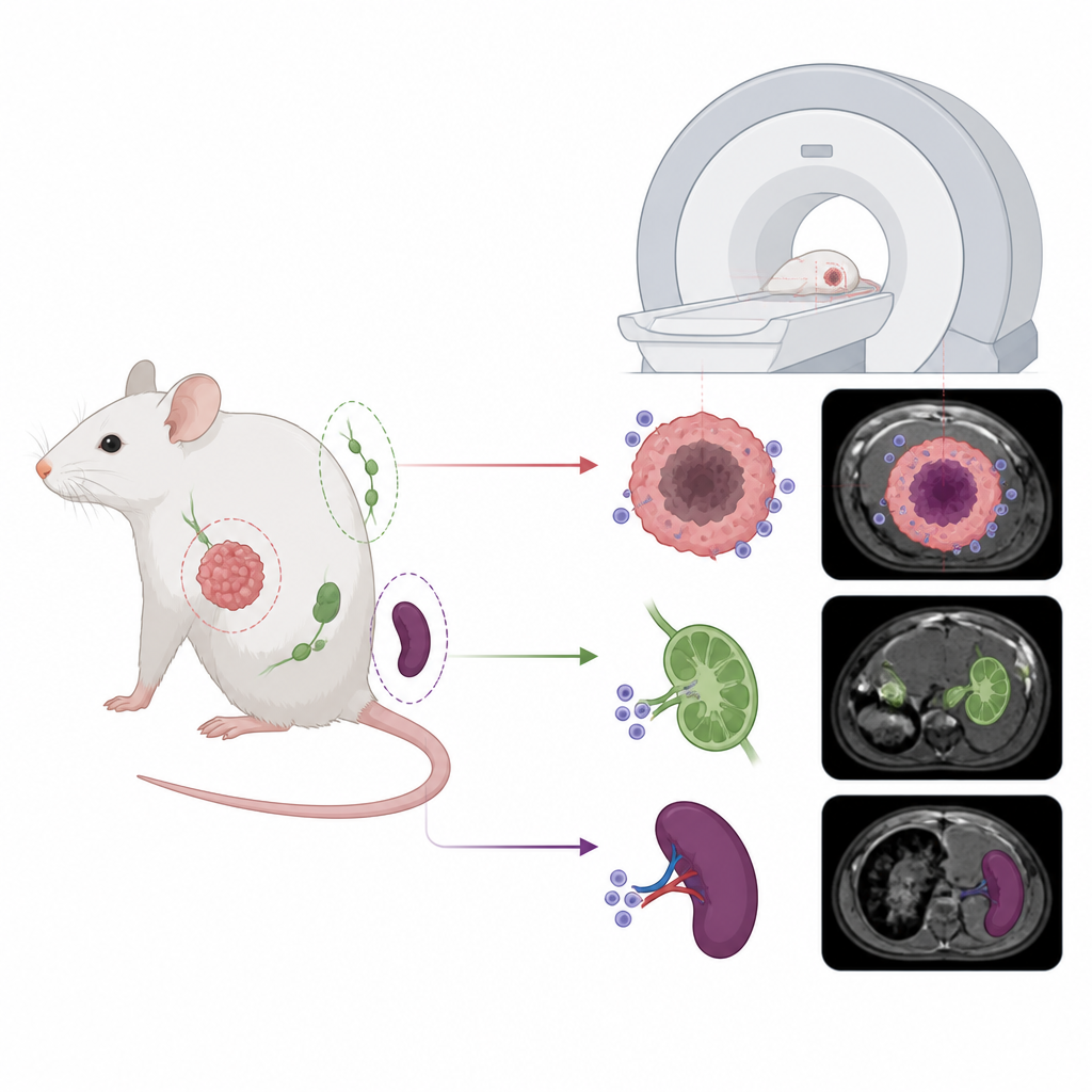
Many solid tumors contain a mix of healthy-looking cancer cells on the outside and dead or dying tissue, called necrosis, in the center. These patterns affect how a tumor grows and how it responds to therapy, but they can be hard to map in detail. The researchers engineered mouse breast cancer cells to carry a “self” reporter gene from the mouse, called mOatp1a1, that makes those cells light up on standard hydrogen MRI after injection of a clinical contrast agent. Because this gene comes from the mouse itself, it is far less likely to trigger immune rejection than foreign optical reporters such as luciferase. With this approach, they could distinguish regions packed with viable engineered cancer cells from areas where cells had died, revealing tumor architecture in three dimensions over time.
Following Immune Cells with a Second MRI Signal
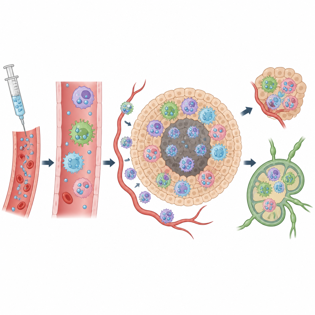
To track immune cells, the team added a second flavor of MRI based on fluorine, using tiny droplets of a perfluorocarbon nanoemulsion injected into the bloodstream. Immune cells naturally take up these droplets, and because normal tissues contain almost no fluorine, any fluorine signal on MRI marks labeled cells. By combining the cancer cell reporter signal with the fluorine signal in the same scan, the scientists could see where immune cells were entering tumors, as well as their presence in the spleen and nearby lymph nodes. Unexpectedly, strong fluorine signal often appeared not just at the tumor edge but also deep inside necrotic cores, hinting that both immune cells and cell debris in dead regions can hold onto the tracer.
Different Organs, Different Immune Mixes
Looking only at images cannot reveal which immune cell types carry the fluorine label, so the researchers used detailed flow cytometry to sort and measure cells taken from tumors, spleens, and lymph nodes. In tumors, fluorine-labeled cells were dominated by myeloid cells such as neutrophils, several kinds of tumor-associated macrophages, and myeloid-derived suppressor cells. In the spleen, the picture was more balanced, with both myeloid cells and lymphocytes like B cells contributing strongly to the signal. In tumor-draining lymph nodes, most labeled cells were T cells and B cells, even though myeloid cells took up more tracer per cell. These organ-specific patterns show that fluorine MRI is not simply a “macrophage meter” but reflects a broad mix of immune players whose makeup depends on the tissue.
What the New Imaging Platform Can Tell Us
Because this dual MRI approach can be used repeatedly in immune-intact mice, it offers a powerful way to follow how tumors evolve and how immune cells move in and out during disease and treatment. The method helps distinguish tumors rich in innate immune cells from those that are relatively poor in such cells, complementing tools that focus on T cells alone. It also highlights that fluorine signal can come from both live immune cells and necrotic pockets, a nuance that is crucial when interpreting preclinical imaging data. Together, tumor-specific hydrogen imaging and immune-resolving fluorine imaging create a more complete map of the tumor neighborhood, which could guide the design and testing of future immunotherapies and improve how animal studies translate to patient care.
Big Picture Takeaway for Patients and Readers
For a lay reader, the key message is that this research brings us closer to watching tumors and immune cells interact in living organisms without surgery. By combining two MRI signals, one tied to living cancer cells and the other to immune cell traffic, scientists can better judge which tumors are packed with immune cells and which are not, and where dead tissue sits inside a mass. While this work is still in mice, it helps explain what fluorine-based MRI really shows and sets the stage for more informative scans that may one day help doctors choose and monitor treatments based on the immune landscape of each person’s tumor.
Citation: McRae, S.W., Lau, J.H., Martinez, F.M. et al. Linking tumor viability and immune infiltration with dual-nucleus MRI in preclinical models. npj Imaging 4, 35 (2026). https://doi.org/10.1038/s44303-026-00158-7
Keywords: tumor microenvironment, immune imaging, fluorine MRI, breast cancer model, myeloid cells