Clear Sky Science · en
Phase-based computational adaptive optics enables artifact-free super-resolution microscopy
Sharper views inside living cells
Modern microscopes can reveal the inner workings of cells in remarkable detail, but the images are often blurred by the very tissues they are trying to probe. This paper introduces a new way to clean up those images after they are taken, using only computation instead of complex moving mirrors or extra sensors. The method promises crisper, more reliable pictures of cells, embryos, and tissues, opening the door for more labs to use ultra‑detailed imaging without buying specialized hardware.
Why microscopes struggle in real tissues
As light travels through a biological sample, it passes regions of different density, such as cell membranes, nuclei, and fluid‑filled spaces. Each change subtly bends the light, much like air turbulence warps the view of stars through a telescope. In microscopes, these bending effects, called optical aberrations, smear out fine details and create misleading patterns and false structures, especially in advanced "super‑resolution" methods that push the limits of clarity. Traditional adaptive optics fight this problem with deformable mirrors and wavefront sensors, but those setups are intricate, expensive, and can expose delicate samples to extra light.
A software-only path to adaptive optics
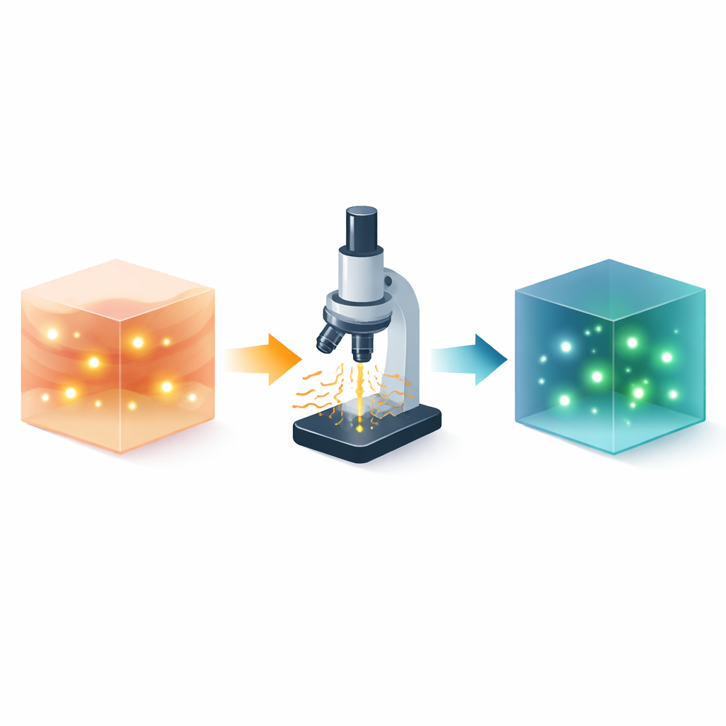
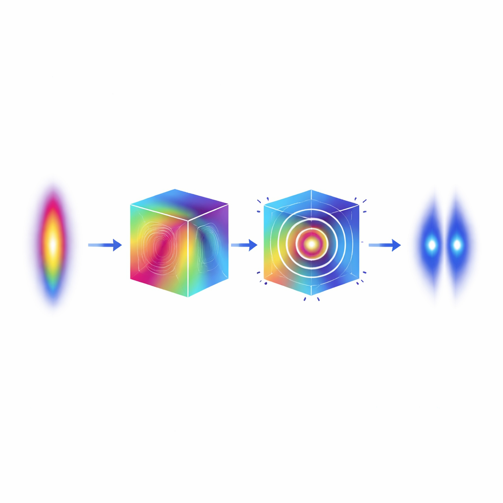
The authors present a purely computational alternative they call phi Computational Adaptive Optics, or ∅CAO. Instead of measuring how the light waves are bent with extra hardware, ∅CAO infers this information directly from the three‑dimensional image stack that the microscope already records. The key insight is that, in 3D, most of the imprint of optical distortion hides not in how bright different parts of the image are, but in the “phase” of the light when the data are converted into frequency space. By carefully adjusting this phase information and then transforming back, the method can undo the bending of light and restore symmetry and sharpness, mimicking what adaptive mirrors do in hardware.
Testing accuracy under tough conditions
To see how well ∅CAO works, the researchers first turned to simulations where the true distortions are known. They created artificial point‑like light sources and thread‑like structures and blurred them with different combinations of common aberrations, including simple lens errors and more complex patterns. ∅CAO was able to recover the correct distortion strengths and restore the images, often in just a few rounds of computation. Even when noise was added so that the signal became faint and grainy, the method still accurately captured the most important kinds of distortion and improved the ability to distinguish nearby details that had previously merged into a single spot.
Bringing clarity back to real biological samples
The team then applied ∅CAO to real microscope data. Fluorescent beads imaged through a standard lens with deliberately mis‑set controls appeared stretched and fuzzy along the viewing direction. After processing with ∅CAO, the beads regained the compact shape seen under optimal settings, and the measured blur matched known lens adjustments. In an especially challenging test, beads viewed through plant leaves—where the tissue causes strong, uneven distortions—were corrected region by region across the field of view, something that is very difficult with hardware alone. Live roundworm embryos imaged in wide‑field mode showed sharper, more distinct chromosomes after correction, and subsequent deblurring algorithms worked better on these cleaned‑up images. The method also greatly improved a popular super‑resolution technique called structured illumination microscopy, removing artificial patterns and ghost structures and restoring fine cellular features in multiple colors.
What this could mean for everyday imaging
Because ∅CAO runs entirely in software and uses only the images that a microscope already produces, it can be added to existing systems without modifying the optics. It shows strong resistance to noise, can handle complex mixtures of distortions, and works on different imaging modes, from standard wide‑field to advanced super‑resolution approaches. The authors argue that focusing on phase information turns a difficult, unstable correction problem into one that reliably converges, and that future machine‑learning tools should build on this principle. While careful selection of image regions is still important and real‑time use will require faster code or hardware acceleration, this phase‑based approach offers an accessible route to adaptive optics for many biology and medical labs.
Clearer pictures for clearer biology
In essence, this work shows that a clever transformation of image data can stand in for complex physical devices, straightening out bent light paths after the fact. By recovering lost detail and removing misleading artifacts across a range of samples—from plant tissues to worm embryos and cultured human cells—∅CAO brings high‑end adaptive optics within reach of standard microscopes. For non‑specialists, the takeaway is simple: better software can make existing microscopes see deeper and more clearly, revealing structures that were always there but hidden in the blur.
Citation: Matsuda, A., Rodriguez-Reza, C.M., Tamada, Y. et al. Phase-based computational adaptive optics enables artifact-free super-resolution microscopy. Commun Eng 5, 75 (2026). https://doi.org/10.1038/s44172-026-00622-7
Keywords: adaptive optics, super-resolution microscopy, image deconvolution, fluorescence imaging, computational imaging