Clear Sky Science · en
Increasing EEG electrode density improves decoding of visual categories and source localization: an exploratory ultra-high-density EEG study
Why packing more sensors on the scalp matters
Every time you look at a face, a body, a household object, or a simple pattern, your brain reacts in just a few hundredths of a second. Electroencephalography (EEG) lets scientists record these fleeting electrical ripples from the scalp, but the picture is often blurry. This study asks a simple question with big implications: if we dramatically increase the number of tiny EEG electrodes over the back of the head, can we “see” visual brain activity more clearly, decode what someone is viewing more accurately, and pinpoint where in the brain it happens?
Seeing brain waves in finer detail
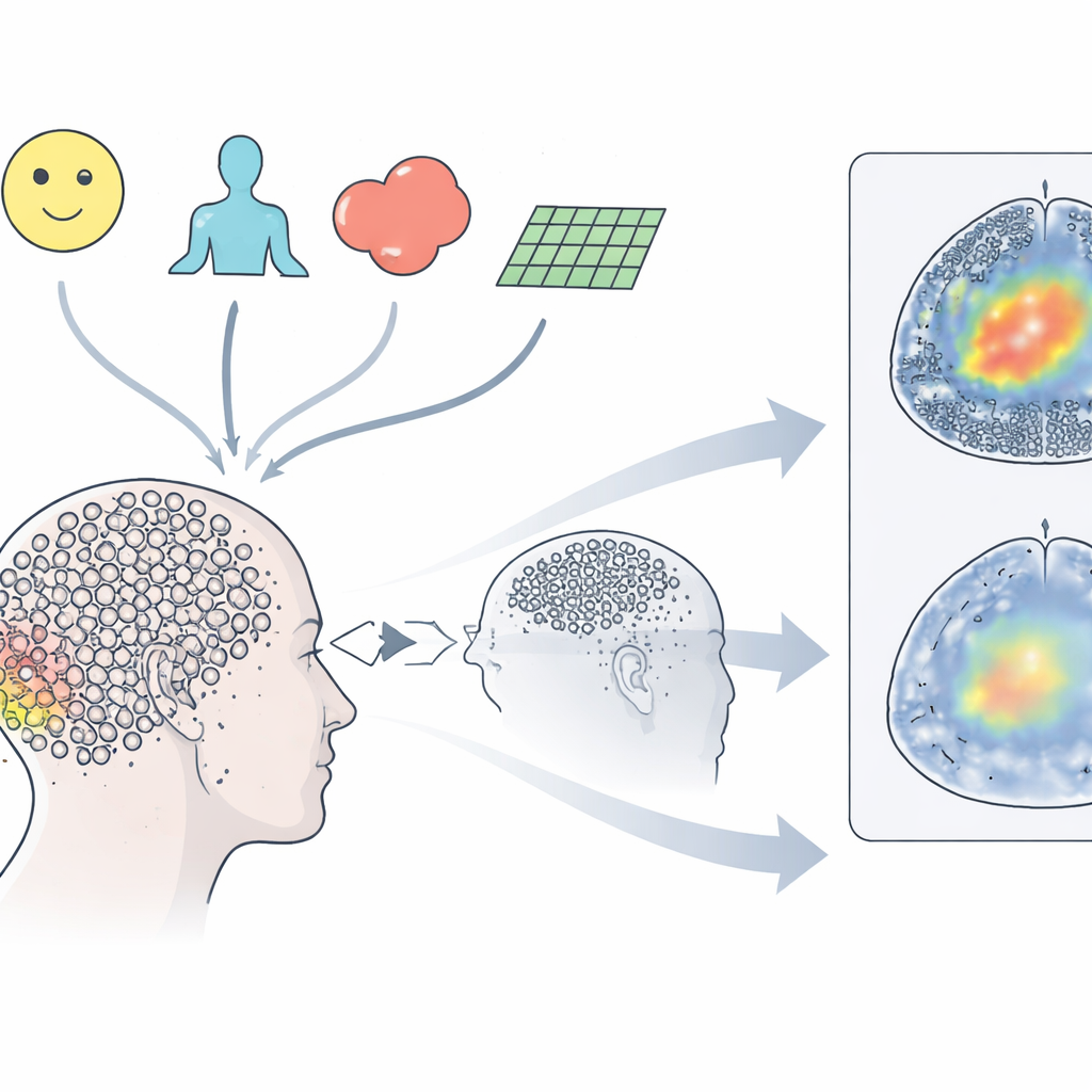
The researchers used an ultra-high-density EEG cap with 512 small electrodes clustered over the occipital region, the area at the back of the head that first processes visual information. Four volunteers viewed hundreds of images from four categories: faces, bodies, everyday objects, and abstract patterns. For each flash of an image, the team measured visual evoked potentials—brief, time-locked waves of electrical activity—that unfold from about one-tenth to half a second after the picture appears. With so many closely spaced sensors, they could create detailed “heat maps” on the scalp showing how activity starts in primary visual areas and then fans out differently for each type of image, for example spreading toward side regions of the brain when faces are shown.
Testing how many sensors are really helpful
To find out whether more electrodes actually improve what can be read from EEG, the team systematically “thinned out” their recordings to mimic standard clinical caps with far fewer sensors. They compared layouts similar to common 10–20 and 10–10 systems with denser variants and finally the full ultra-dense setup. Using a straightforward statistical classifier, they tried to guess on each individual trial which of the four categories a person was seeing. Accuracy climbed steadily with sensor density: traditional layouts averaged just under 60 percent correct, while the ultra-dense grid reached about 73 percent, with some participants exceeding 76 percent. Crucially, packing electrodes closer together mattered more than simply spreading them over a wider area of scalp, suggesting that fine spatial sampling over the key visual region is especially valuable.
Following the brain’s timing more precisely
Beyond overall accuracy, the authors examined when in time the brain’s patterns first carried enough information to distinguish one category from another. They trained their classifier at one moment after stimulus onset and tested whether it could generalize to other time points, building a “temporal map” of decodability. With denser electrodes, decoding not only became more accurate but also kicked in earlier—around 70 milliseconds after the image appeared, peaking near 150–200 milliseconds. This indicates that better spatial sampling on the scalp sharpens the apparent timing of brain events as well, reducing blurring caused by electrical signals spreading through the head.
Tracing signals back into the brain
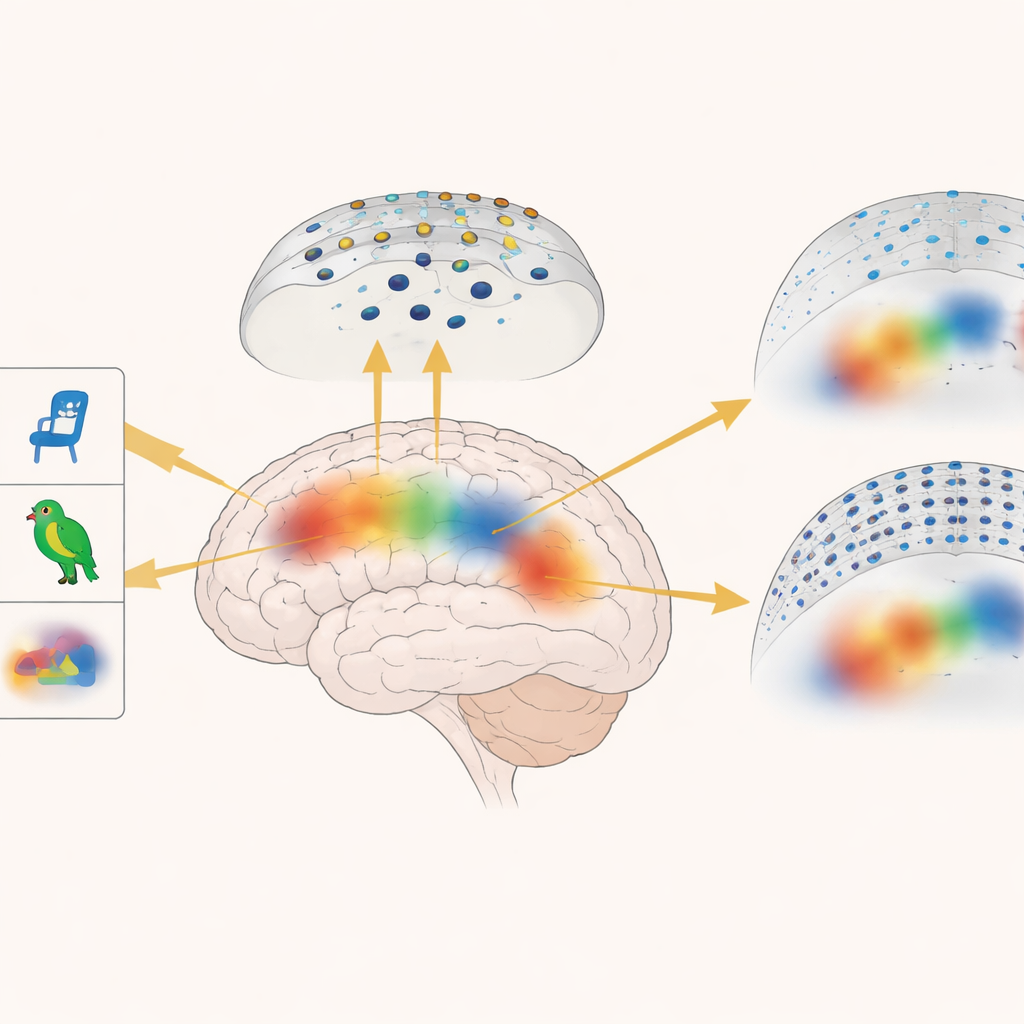
High electrode density also improved the next step: estimating which brain regions generated the observed scalp signals. Using each participant’s MRI scan and established source-localization algorithms, the team reconstructed where activity likely originated inside the brain. Early responses for all categories clustered in the primary visual cortex at the back of the brain. Later, activity moved into regions along the underside of the temporal lobes that are known to support recognition of objects and faces. For faces in particular, the method pinpointed a response around 170 milliseconds in the fusiform gyrus, a region long linked to face perception. When the same analysis was repeated with sparser, simulated layouts, these internal activation patterns became blurrier and less focal, underscoring the added value of ultra-dense recordings.
From basic vision science to future applications
Although the study involved only four volunteers and focused on a limited patch of scalp, it demonstrates that packing many small electrodes into a key region can make EEG both more informative and more precise. Denser layouts boosted the ability to tell what kind of image someone was viewing, clarified how activity spreads across the scalp, and sharpened estimates of where and when specific brain regions—such as the fusiform gyrus for faces—come online. For everyday readers, the takeaway is that upgrading EEG from a coarse grid to an ultra-fine mesh could transform it from a rough stethoscope of the brain into a more detailed sensor, with potential benefits for brain–computer interfaces, diagnostics, and research on how we see and recognize the world.
Citation: Schreiner, L., Sieghartsleitner, S., Kapeller, C. et al. Increasing EEG electrode density improves decoding of visual categories and source localization: an exploratory ultra-high-density EEG study. Commun Eng 5, 59 (2026). https://doi.org/10.1038/s44172-026-00611-w
Keywords: ultra high density EEG, visual perception, brain decoding, source localization, brain computer interface