Clear Sky Science · en
hERG epitope mimic-decoy peptide corrects autoimmune-long QT syndrome in guinea pigs
Why this matters for heart health
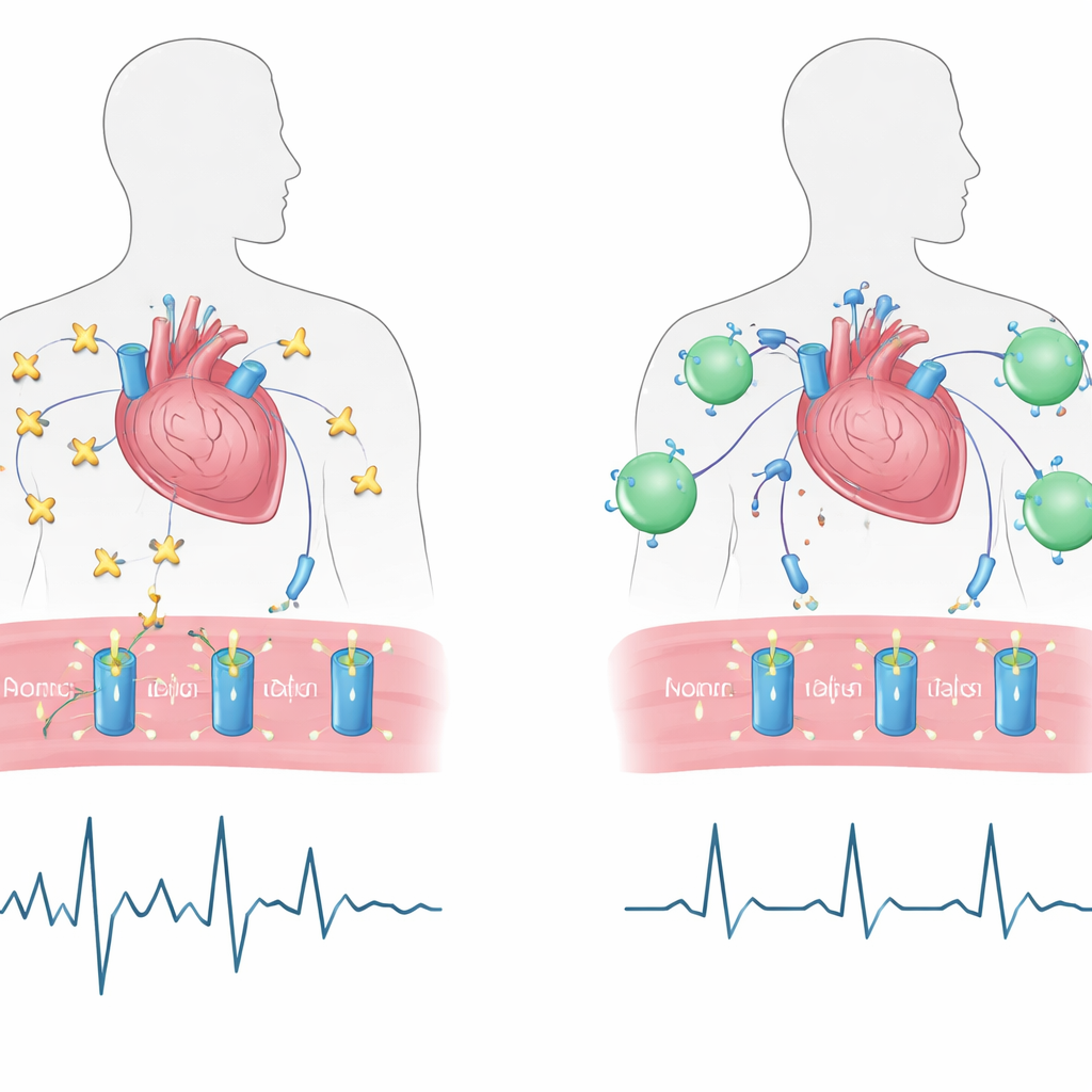
Many people carry immune proteins that quietly attack their own bodies without obvious symptoms. Some of these wayward antibodies can subtly sabotage the heart’s electrical system, stretching out the time each heartbeat takes to reset. That hidden glitch, called long QT, can set the stage for dangerous rhythm disturbances and sudden cardiac death. This study explores a new strategy: a custom-built “decoy” protein, MDP4, designed to lure away harmful antibodies before they can disrupt the heart’s electrical channels, at least in guinea pigs.
When the immune system misfires on the heart
The work focuses on a form of acquired long QT syndrome driven by autoantibodies against a protein called Ro/SSA-52kD. These antibodies, originally made in some autoimmune diseases but also present in a fraction of otherwise healthy people, can latch onto a crucial potassium channel in heart cells known as hERG. That channel helps the heart’s electrical signal recover after each beat. When antibodies clamp onto a small region on hERG, they weaken the current (IKr) that flows through it, stretching the QT interval seen on an electrocardiogram (ECG) and raising the risk of dangerous arrhythmias such as Torsades de Pointes. Epidemiologic studies suggest that people with these antibodies have roughly double the risk of significant QT prolongation and serious rhythm problems, making this a wider public health concern than once thought.
A designer decoy built from channel parts
To counter this problem, the researchers engineered a hybrid protein called monobody decoy peptide 4, or MDP4. They started from a short stretch of the hERG channel’s outer pore region—the very sequence the harmful antibodies recognize, nicknamed “peptide 4.” On its own, this linear fragment would be unstable and prone to clumping in the body. The team therefore fused it to a compact, well-behaved human protein scaffold known as a fibronectin monobody, which is already considered non-immunogenic and has been used safely in clinical trials. Using three-dimensional protein modeling and energy calculations, they predicted how the fused peptide would fold so that the hERG-like segment would be displayed in a lifelike shape, ready to attract the antibodies in place of the real channel.
Putting the decoy to the test in animals
The authors then used an established guinea pig model of autoimmune long QT syndrome. Animals were immunized with the Ro/SSA-52kD antigen over two weeks, prompting them to produce anti-Ro/SSA-52kD antibodies. As expected, their heart tracings showed a progressive lengthening of the rate-corrected QT interval (QTc). After confirming robust antibody levels, one group of guinea pigs received injections of MDP4, a second group received a control monobody lacking the hERG segment, and a third group was left untreated. In untreated and control animals, QTc remained significantly prolonged. In contrast, animals given MDP4 saw their QTc return to baseline values within about 15 days, without notable changes in heart rate, PR interval, or QRS width. Blood tests confirmed that MDP4 circulated in the bloodstream and that antibodies from immunized animals recognized the hERG-like peptide, consistent with the decoy concept.
How the decoy restores the heart’s electrical reset
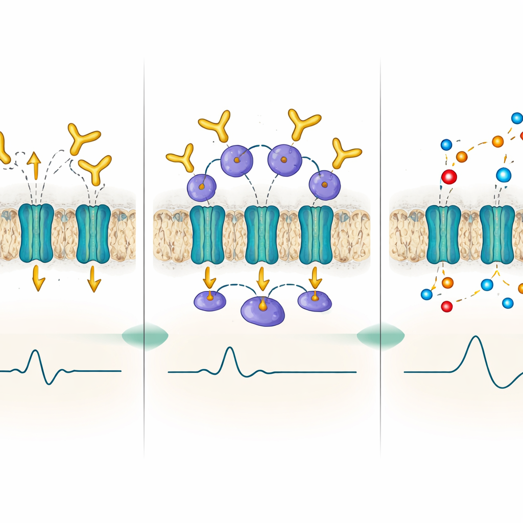
To probe the mechanism, the team moved to isolated cells. They first confirmed that MDP4 itself does not significantly change the potassium current in cells engineered to express hERG or in normal guinea pig heart cells, suggesting it is not directly blocking or activating the channel. Next, they exposed heart cells to IgG antibodies from a patient with long QT and Torsades de Pointes whose blood contained strong anti-Ro/SSA-52kD activity. As shown in earlier work, these antibodies reduced the hERG-related current (IKr) and prolonged the duration of the action potential, mirroring long QT. When MDP4 was added after the damaging antibodies, IKr returned to near-normal levels and the prolonged action potential shortened back toward baseline, indicating that the decoy could rescue function even after injury had begun. When cells were pretreated with MDP4 before antibody exposure, the current remained essentially unchanged, implying that the decoy can also act preventively by soaking up antibodies before they reach the channel.
Promise and limits of a precision decoy
This study shows that a rationally designed decoy protein can correct immune-driven electrical abnormalities in the heart of an animal model, without obvious direct side effects on the key channel. Because MDP4 mimics only a tiny, disease-relevant patch of hERG, the approach is highly targeted: it aims not to blunt the entire immune system but to intercept one harmful interaction. The authors note that more work is needed to assess long-term safety, immune reactions to the decoy itself, and effects on other heart currents, as well as to determine dosing and effectiveness in humans. Still, as a proof of principle, MDP4 demonstrates that it may be possible to reverse certain antibody-mediated rhythm disorders by competitively blocking autoantibodies at their source, potentially opening a path to similar precision decoys for other autoimmune heart channel diseases.
Citation: Cupelli, M., Ginjupalli, V.K.M., Reisqs, JB. et al. hERG epitope mimic-decoy peptide corrects autoimmune-long QT syndrome in guinea pigs. Commun Med 6, 245 (2026). https://doi.org/10.1038/s43856-026-01508-7
Keywords: autoimmune long QT syndrome, hERG potassium channel, decoy peptide therapy, cardiac arrhythmia, autoantibodies