Clear Sky Science · en
Predicting categorical and continuous Alzheimer’s disease outcomes from a single MRI scan
Why this study matters to families and doctors
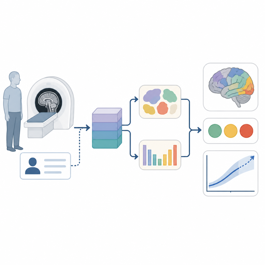
Alzheimer’s disease slowly steals memory and thinking, but tracking who will decline, and how fast, usually demands long, stressful testing sessions and specialized scans. This study asks a simple but powerful question: can a single routine brain scan, plus basic background information, forecast not only whether someone has Alzheimer’s, but also how their thinking will change over the next few years? The answer, from a new artificial intelligence system, is cautiously encouraging.
A new way to read a brain scan
The researchers focused on magnetic resonance imaging, or MRI, which is already common in hospitals and clinics. Traditionally, MRI is used to look for visible signs of stroke, tumors or shrinkage in certain brain areas. Here, the team instead fed three-dimensional MRI images into deep learning models that can spot subtle patterns far beyond what the human eye can see. They combined this with a few simple details about each person, such as age, sex, education level and marital status, avoiding expensive blood tests, genetic panels or specialized imaging like positron emission tomography.
From one scan to many answers
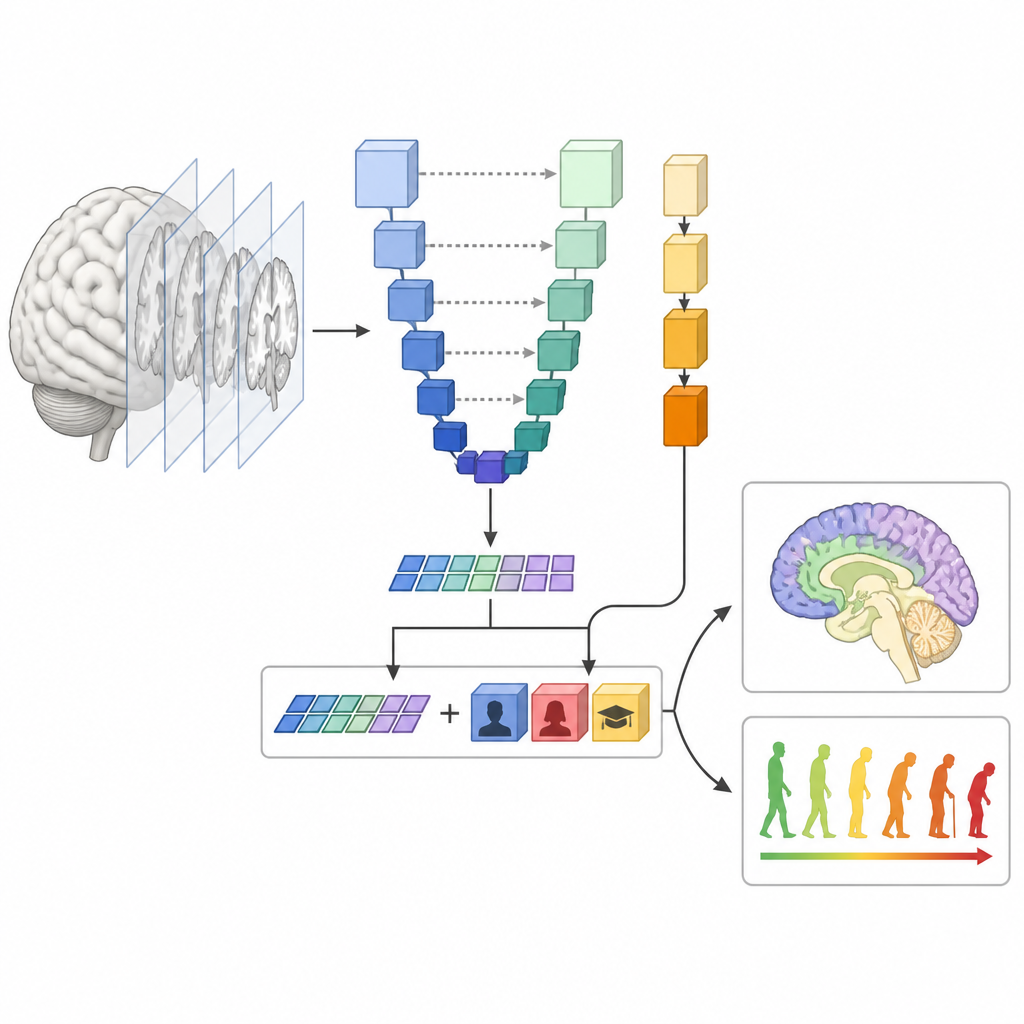
The heart of their approach is a multitask system that learns to solve several related problems at once from the same scan. One branch of the model chops the brain image into three main tissue types: gray matter, white matter and fluid-filled spaces. Another branch uses the same internal features to judge whether the person falls into a healthy, mildly impaired or Alzheimer’s group. At the same time, the system learns to estimate a widely used cognitive test score, called ADAS-Cog, which summarizes memory and thinking ability on a continuous scale. Training these tasks together helps the model discover brain features that matter for both anatomy and cognition.
Teaching computers with many brains
To build and test the system, the team used thousands of MRI scans and cognitive scores from large public studies of aging and memory. They compared several model designs, including a custom-built network and a very large, general-purpose medical imaging model that they carefully adapted to brain scans. They also combined their image-based models with a separate tree-like algorithm that only sees the simple background information and summary tissue volumes. In the end, they blended the predictions from these parts into an ensemble that gives more weight to whichever component does better on each task.
How well did it work?
On people held back from training, the final ensemble models were able to segment brain tissue with accuracy on par with popular, slow research tools, but in a much more streamlined way. They correctly distinguished Alzheimer’s from non-Alzheimer’s cases in more than nine out of ten participants. Most strikingly, they estimated current thinking scores with relatively small average errors, and these estimates stayed meaningful when the model tried to forecast scores up to three years into the future using only the first scan. The system also held up reasonably well on an entirely separate group of generally healthy adults scanned at different centers, though performance dipped somewhat, highlighting how differences in scanners and populations still matter.
Peeking inside the black box
The authors went beyond raw accuracy and asked which inputs the model relied on most. When they disrupted small patches of the MRI image, performance dropped most in regions already known to be important in Alzheimer’s, such as the hippocampus and nearby temporal and parietal areas. Overall, the brain images themselves contributed more to predictions than background information, but age, education and marital status still played clear roles. The team also showed that traditional summaries of brain volume, extracted with time-consuming software, could not match the accuracy of their direct image-based approach.
What this could mean for patients
For patients and clinicians, these findings suggest that a standard brain scan, interpreted by a carefully designed artificial intelligence system, could one day provide a quick snapshot of both current thinking ability and likely future decline. Such tools might help decide who needs closer monitoring, who could benefit from specialist evaluation or trial enrollment, and how to plan care before symptoms become severe. The work does not replace existing tests or prove that MRI alone is sufficient, but it offers a practical step toward using information already collected in everyday clinics to make dementia care more proactive and more personalized.
Citation: Ma, D., Pabalan, C., Rajagopal, A. et al. Predicting categorical and continuous Alzheimer’s disease outcomes from a single MRI scan. Nat Aging 6, 1121–1137 (2026). https://doi.org/10.1038/s43587-026-01121-2
Keywords: Alzheimer’s disease, brain MRI, cognitive decline, deep learning, prognosis