Clear Sky Science · en
Soft-interfaced liquid crystal microfluidics can probe the rigidity of lipid vesicles
Watching Cell Membranes in Action
Many serious diseases, from cancer to neurodegeneration, are linked not just to what is inside our cells, but to how stiff or flexible their outer membranes are. Yet measuring these tiny mechanical changes is slow and technically demanding. This paper presents a new way to "see" membrane stiffness in real time by using a special kind of fluid, a liquid crystal, inside a microfluidic chip. Changes in how model cell membranes fuse with this soft interface cause dramatic optical shifts, offering a potential early-warning tool for membrane-related diseases.
A Soft Window into Cell-Like Membranes
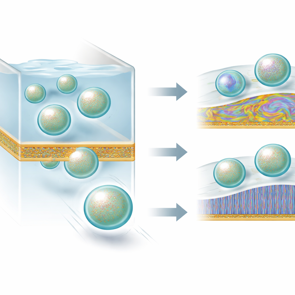
The researchers build on the unusual properties of liquid crystals—the same materials used in LCD screens. In their nematic phase, these fluids have molecules that tend to point in the same direction, making them very sensitive to what happens at their surface and easy to read optically under crossed polarizers. At the boundary between a liquid crystal and water, the orientation of the liquid crystal molecules changes when a film of lipid molecules—similar to those in cell membranes—forms on that interface. Depending on whether the lipid layer covers only patches or the entire surface, the liquid crystal appears bright or dark, effectively turning invisible molecular events into visible patterns.
Microfluidic Channels as Test Tracks
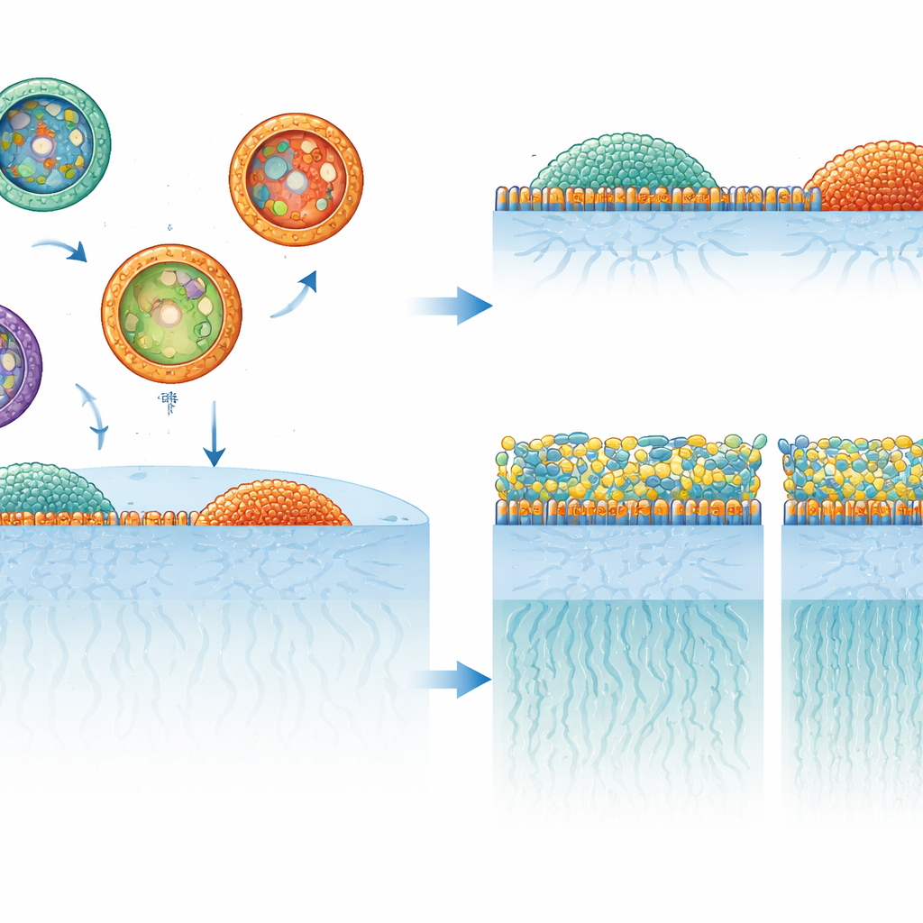
To exploit this sensitivity, the team created three experimental setups: flat liquid crystal films held in tiny grids, microscopic liquid crystal droplets floating in water, and, most importantly, microfluidic channels where a liquid crystal and an aqueous solution flow side by side. In the channels, the two fluids form a stable, horizontal interface. When lipid vesicles—small, membrane-bound spheres mimicking cell membranes—are carried along by the flow, some of them collide with the interface and fuse, spreading their lipids into a thin film. The liquid crystal beneath responds by rearranging its internal order, producing bright and dark domains that can be followed over time and along the channel length.
How Stiffness and Additives Change Fusion
The authors systematically compared vesicles made from different phospholipids that are either fluid or gel-like at room temperature. Soft vesicles formed from DLPC or DOPC fused readily with the liquid-crystal interface, quickly generating extensive lipid coverage and strong changes in optical appearance. In contrast, very rigid vesicles made from egg sphingomyelin barely fused at all, and DPPC vesicles, also fairly rigid, fused only when subjected to shear—either by flow in the microchannel or vigorous mixing in droplet experiments. By adding guest molecules that soften membranes (a surfactant) or stiffen them (a ceramide), the researchers could speed up or almost completely halt fusion, showing that the optical response directly reflects vesicle mechanical properties rather than simple diffusion.
Cholesterol’s Surprising and Lipid-Specific Role
Cholesterol, a key component of real cell membranes, has a particularly complex influence on membrane stiffness. Using their liquid-crystal platform, the team tracked how adding cholesterol to different lipids altered fusion behavior. For DLPC and DOPC, increasing cholesterol first made vesicles harder and less likely to fuse, reducing interfacial coverage, but beyond about 50 percent cholesterol the trend reversed and fusion sped up again, suggesting a softening of highly cholesterol-rich vesicles. With DPPC, fusion steadily became more difficult as cholesterol increased, consistent with a monotonic stiffening. Egg sphingomyelin behaved in the opposite way: initially too rigid to fuse, its vesicles began to fuse once enough cholesterol was added, implying that cholesterol was actually softening these already stiff membranes. Independent surface rheology measurements on lipid monolayers supported these lipid-specific trends in mechanical behavior.
From Optical Patterns to Possible Diagnostics
By tying the speed and extent of vesicle fusion to how the liquid crystal interface looks under the microscope, this work turns subtle mechanical properties of membranes into simple, visible signals. Because vesicles similar to those studied here are shed by real cells and circulate in body fluids, the same strategy could eventually help detect early changes in membrane rigidity associated with cancer, metabolic disorders, or neurodegenerative disease. The microfluidic liquid-crystal platform offers a continuous, label-free way to monitor how soft or stiff membrane-bound particles are, opening a path toward compact diagnostic devices that read cell health from shifting optical textures rather than complex biochemical labels.
Citation: Dedeoglu, C., Bukusoglu, E. Soft-interfaced liquid crystal microfluidics can probe the rigidity of lipid vesicles. Commun Mater 7, 120 (2026). https://doi.org/10.1038/s43246-026-01128-7
Keywords: liquid crystal microfluidics, lipid vesicle rigidity, cholesterol and membranes, membrane mechanics sensing, extracellular vesicle diagnostics