Clear Sky Science · en
Ultra-low field 13C MRI of hyperpolarized pyruvate
Seeing Disease Before It Changes Shape
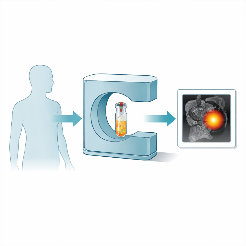
Many illnesses, especially cancer and heart disease, quietly change how our cells use energy long before organs look abnormal on a scan. Today’s hospital MRI machines mostly show anatomy: size, shape, and structure. This paper explores a way to turn MRI into a metabolic camera that could be cheaper, more portable, and able to detect disease earlier by watching how a simple fuel molecule, pyruvate, is processed in the body.
Why Cell Fuel Matters
Pyruvate is a small molecule that sits at a crossroads of cellular metabolism, helping decide whether fuel is burned cleanly with oxygen or rapidly fermented, a tendency that often shifts in cancer and other diseases. Doctors already test pyruvate’s behavior in patients using “hyperpolarized” MRI, which boosts the faint signal of carbon atoms so strongly that their metabolic fate can be tracked in real time. But the current technology that creates these bright signals is huge, costly, and slow, restricting such scans to a handful of elite centers worldwide. To make metabolic imaging a practical tool for everyday care, the machinery must become faster, cheaper, and easier to install.
A Simpler Way to Supercharge MRI Signals
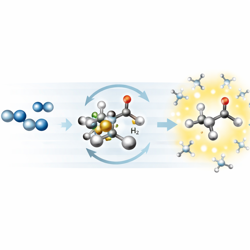
The researchers build on an emerging method called SABRE, which borrows order from a special form of hydrogen gas and hands it to a target molecule without permanently changing its chemistry. In their variant, called SLIC SABRE, a carefully tuned radio wave “locks” the spins of the atoms at just the right frequency so that this order flows efficiently into carbon-13 atoms in pyruvate. Unlike the conventional approach that needs extremely cold temperatures and very strong magnets, this method works at a magnetic field thousands of times weaker than a hospital MRI and uses hardware that can be built for a fraction of the cost. In this study, they keep the entire process inside an open, ultra-low-field scanner operating at just 6.5 millitesla, about one-thousandth of a typical clinical system.
Making Metabolism Glow at Ultra-Low Field
Inside the small scanner, the team bubbles parahydrogen gas through a solution containing pyruvate and a metal-based catalyst. Under the right conditions of temperature, gas flow, and radio-wave strength, pyruvate molecules repeatedly bind to and detach from the catalyst while the hydrogen donates its hidden order. Within about 10 seconds, the carbon-13 signal from pyruvate is boosted by more than a million times compared with what thermal equilibrium would provide at this weak field, reaching polarization levels of around 3 percent. That jump in signal is large enough to not only detect pyruvate easily, but also to resolve subtle frequency differences that separate forms of pyruvate and reveal whether it is free in solution or still bound to the catalyst.
Turning Hyperpolarization into Images
Signal alone is not enough; it must be turned into pictures. The authors adapt and invent MRI pulse sequences that tap into this bright but short-lived magnetization. In a “single-shot” approach, they generate hyperpolarization once, quickly store it, and then read it out with a three-dimensional imaging sequence tuned for ultra-low field. This produces clear 3D images of a small sample of hyperpolarized pyruvate in just seconds, with signal strong enough to map its distribution. In a second, “multi-shot” approach, they repeatedly re-hyperpolarize the sample before each line of data is collected, effectively renewing the signal and even capturing how gas bubbles move through the tube. Alongside these images, the team records high-resolution spectra at the same low field, showing that they can distinguish different carbon positions in pyruvate and identify fine structure in the signals that will later help separate pyruvate from its metabolic products.
From Lab Bench to Bedside Possibilities
Although these experiments are performed in a test tube, they outline a realistic route toward affordable metabolic MRI. By marrying fast, inexpensive hyperpolarization based on parahydrogen with ultra-low-field scanners that are small and flexible enough to be placed in regular hospital rooms or even remote clinics, the work points to a future in which metabolic imaging could become routine rather than rare. The study shows that, even at very weak magnetic fields, it is possible to create bright carbon-13 signals, form detailed three-dimensional images, and separate chemical fingerprints well enough to track metabolism. If translated to living subjects, such systems could help doctors see dangerous metabolic shifts in tumors, heart muscle, or the brain long before anatomy changes, opening the door to earlier diagnosis and more personalized, responsive treatment.
Citation: Boele, T., McBride, S.J., Pike, M. et al. Ultra-low field 13C MRI of hyperpolarized pyruvate. Commun Chem 9, 169 (2026). https://doi.org/10.1038/s42004-026-01971-2
Keywords: metabolic MRI, hyperpolarized pyruvate, ultra-low-field imaging, parahydrogen SABRE, personalized medicine