Clear Sky Science · en
A fluorescent probe under the microscope showing dual recognition of B-DNA and G-quadruplex DNA
Why a Color-Changing DNA Marker Matters
Inside every cell, DNA folds into shapes that influence how genes are turned on or off. Being able to see these shapes in real time could transform how we study cancer, aging, and gene regulation. This paper explores a special fluorescent molecule that glows in different colors when it binds to two major DNA forms, and uses advanced computer simulations to explain how one tiny probe can both recognize and color-code these distinct DNA structures.
A Shape-Shifting Light for DNA
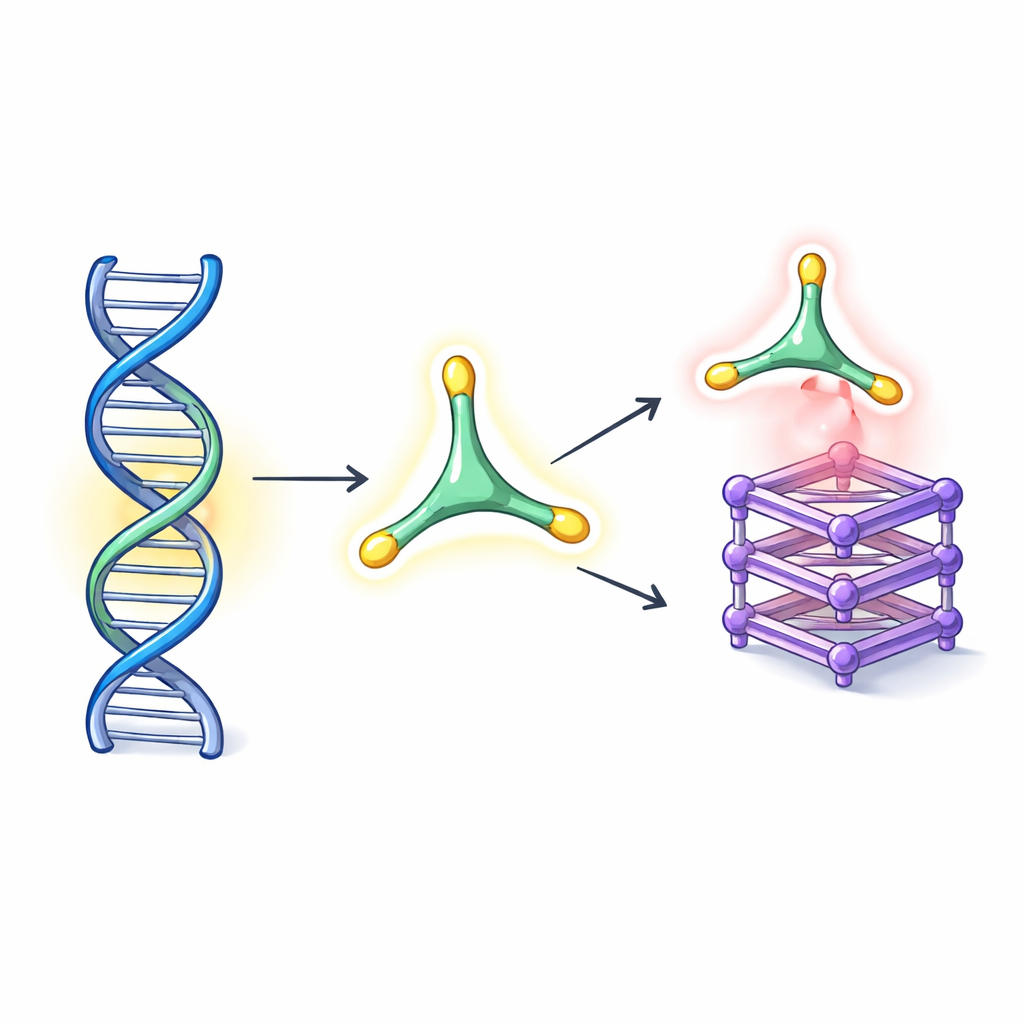
The study focuses on a star-shaped dye called QCy(MeBT) 3, designed to act as a "switch-on" light: it glows much more strongly when it latches onto DNA. Remarkably, experiments had shown that this single molecule brightens in one color when attached to the familiar double-helix form of DNA (B-DNA) and in a more red-shifted color when bound to G-quadruplex DNA, a compact structure formed by stacks of guanine-rich layers. These G-quadruplexes appear in telomeres and gene promoters and are considered attractive targets for anticancer strategies. However, no one really knew why this one probe could both distinguish between these DNA shapes and report on them with distinct colors.
Following Molecular Motion in Silico
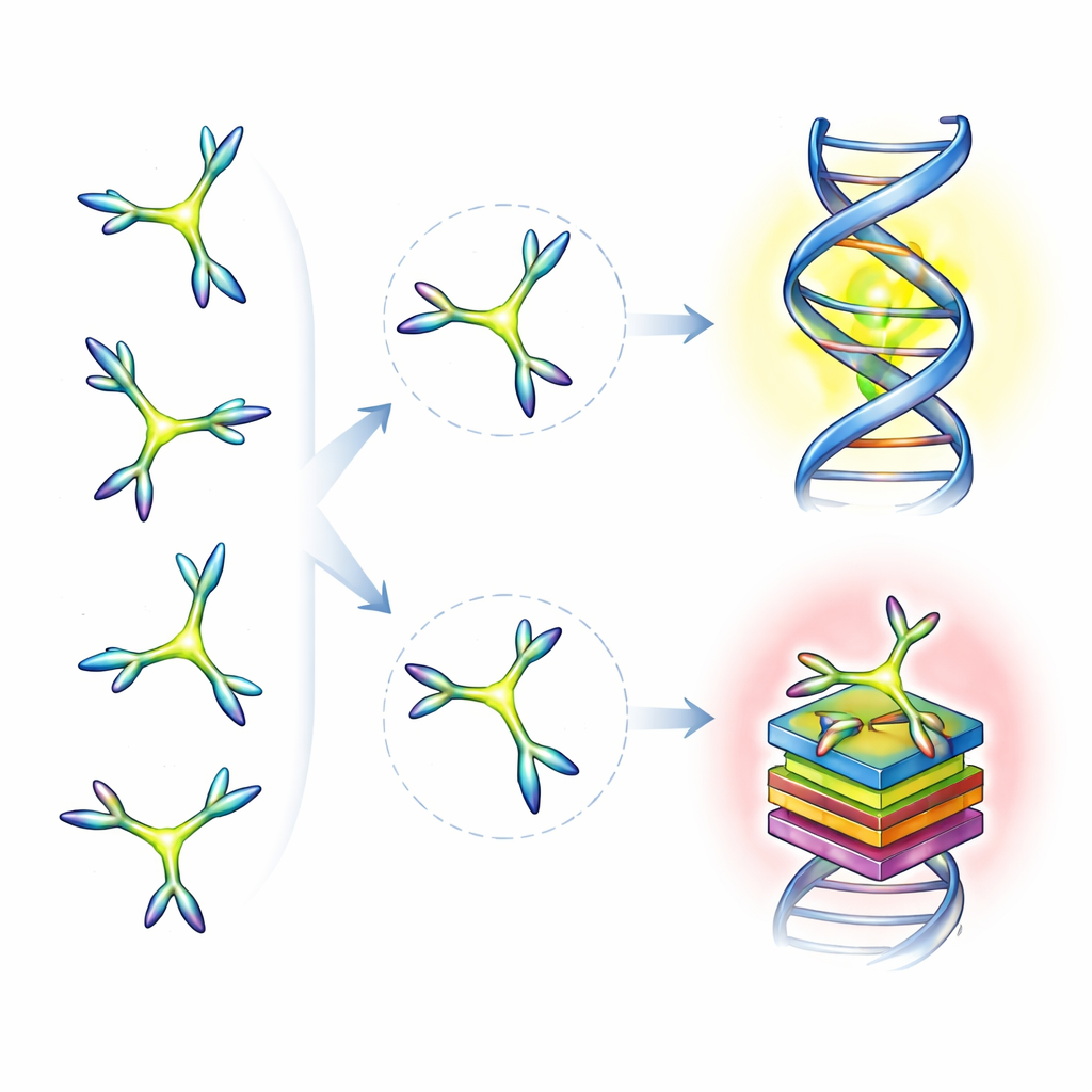
To uncover the mechanism, the authors built a multistep computational protocol that mirrors, in silico, what happens in a cell-like environment. They first mapped all the ways the three arms of the star-shaped probe can twist relative to its central core. Each arm can adopt one of four torsional positions, giving rise to 32 chemically distinct overall shapes, or conformations. Quantum chemistry calculations showed which conformations are most stable in water, and long molecular dynamics simulations then tracked how each version of the molecule moves and interconverts over hundreds of nanoseconds, both free in solution and when near B-DNA or G-quadruplex DNA.
Different DNA, Different Favorite Shapes
The simulations revealed that the probe’s flexibility is not just a minor detail—it is the key to its double life. In water, one particular conformation dominates, but this "resting" shape is not the one that actually binds DNA. Instead, as the arms twist, a few specific conformations act as the best fit for each DNA structure. For G-quadruplexes, only two conformations account for most of the binding, while two different conformations almost entirely dominate binding to B-DNA. Despite this selective matching, the overall binding strength to both DNA forms is similarly high, on par with some well-known G-quadruplex-stabilizing drug candidates, suggesting that QCy(MeBT) 3 is not just a reporter but can also help stabilize these DNA structures.
How Binding and Color Are Linked
Once the preferred conformations and binding poses were identified, the team used hybrid quantum mechanics/molecular mechanics methods to compute absorption and fluorescence spectra and compare them with experiments. They found that the probe binds to G-quadruplexes mainly by stacking its flat aromatic core on the top guanine layer, combining electrostatic attraction and van der Waals contacts. In B-DNA, the same core and two arms slide into the minor groove, guided largely by electrostatic attraction to the negatively charged backbone, while the third arm largely hangs free. Crucially, the conformations that best recognize each DNA type are also the ones that dominate the light-absorption and light-emission behavior. Depending on which conformation is excited at a given wavelength, the intensity and color of the emitted light shift, with the G-quadruplex-bound conformers tending toward deeper red emission than those favored by B-DNA.
Color Coding DNA Shapes by Design
The central takeaway for a non-specialist reader is that the probe’s own shape, not just the DNA’s shape, decides what color we see. The work shows that each flexible conformation of the molecule has its own preferred DNA partner and its own characteristic emission color, and that the redder glow seen with G-quadruplex DNA can be predicted from how those conformations behave in water alone. This insight suggests a powerful design rule: by tuning the internal flexibility and locking in certain conformations, chemists could deliberately craft fluorescent markers that either recognize multiple DNA structures with different colors or selectively target a single DNA topology. Such rationally designed, color-coded probes could become valuable tools for imaging genome organization, tracking cancer-related DNA structures, and coupling diagnosis with therapy in future "theranostic" applications.
Citation: Gramolini, L., López-Corbalán, R., Marazzi, M. et al. A fluorescent probe under the microscope showing dual recognition of B-DNA and G-quadruplex DNA. Commun Chem 9, 164 (2026). https://doi.org/10.1038/s42004-026-01960-5
Keywords: fluorescent DNA probe, G-quadruplex, B-DNA, molecular dynamics, dual-color imaging