Clear Sky Science · en
High-energy resolution X-ray spectroscopy reveals bonding characteristics of La3+ homologues of actinium radiopharmaceuticals
Why this research matters for cancer care
Targeted alpha therapy is an emerging form of cancer treatment that uses tiny bursts of intense radiation to destroy tumors while sparing healthy tissue. A key challenge is building drug molecules that can grip radioactive metals such as actinium tightly enough to carry them safely through the body. Because actinium is rare and hard to handle, this study turns to its non-radioactive cousin lanthanum to work out, in atomic detail, how these metals bond to medical carrier molecules. The team shows that advanced X-ray techniques can reveal how strongly the metal and its surrounding atoms share electrons, information that is crucial for designing safer and more effective radiopharmaceuticals.
Metal cages for powerful medicines
In targeted alpha therapy, actinium atoms are attached to organic “chelators” that act as cages, guiding the radioactive payload to cancer cells and holding it in place while it decays. The stability and behavior of these cages depend on very small differences in how the metal ion binds to oxygen and nitrogen atoms in the chelator. Directly studying actinium is difficult, so the researchers use lanthanum, which has a similar charge and size but is much easier to work with. They focus on several medically important ligands, including the widely used DOTA framework, the fast-binding MACROPA chelator, the clinically applied PSMA-617 construct for prostate cancer, simple water molecules, and a common pH buffer called TRIS. 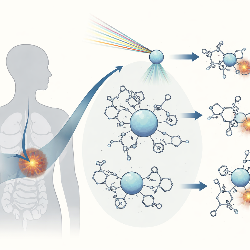
Seeing bonds with sharpened X-ray eyes
To probe these bonds, the team harnesses two high-resolution X-ray techniques that act like finely tuned cameras for electrons. In core-to-core resonant inelastic X-ray scattering (CC-RIXS), one X-ray photon excites an inner electron of lanthanum and a second photon is emitted as the system relaxes; the detailed map of incoming versus outgoing energies encodes how electrons occupy different shells around the ion. High-energy-resolution X-ray absorption near-edge structure (HR-XANES) then zooms in on the sharp rise in absorption at the so-called L2 edge of lanthanum, where small shoulders and pre-edge features betray subtle mixing between metal and ligand orbitals. Together with sophisticated quantum-chemical calculations, these measurements allow the researchers to separate the roles of two key orbital types—compact 4f and more extended 5d shells—and to quantify how much they participate in bonding.
Translating spectra into bonding strength
The spectra reveal two complementary yardsticks for bond character. In the CC-RIXS maps, the separation between a pair of weak pre-edge signals changes systematically among the different complexes. Theory shows that this gap shrinks when the 4f shell participates more in bonding, a phenomenon related to the so-called nephelauxetic effect, where electron–electron repulsion is reduced as orbitals spread out and share density with surrounding atoms. HR-XANES provides a second gauge: the energy distance between a faint pre-edge peak and the main absorption feature reflects how the 4f and 5d levels shift under the influence of the ligands. Larger separations correspond to more ionic, less shared bonding, while smaller separations signal increased covalency, where electrons are more strongly shared between the metal and its neighbors.
Ranking medical chelators at the atomic level
By applying these spectral metrics across all complexes, the authors organize the ligands along a scale from mostly ionic to more covalent bonding. Simple lanthanum in water behaves largely ionically, with little electron sharing beyond basic electrostatics. DOTA and MACROPA, in contrast, induce measurable covalency, but through slightly different channels: MACROPA enhances interaction with 4f orbitals, while DOTA more strongly perturbs the 5d shell. The clinically used PSMA-617 chelator shows a covalency level similar to DOTA, consistent with its robust performance in therapy. Additional quantum-chemical analyses, which decompose interaction energies and track how much electron density is actually shared, support these trends and show that even when bonds look similar in distance, their electronic nature can differ in subtle but important ways. 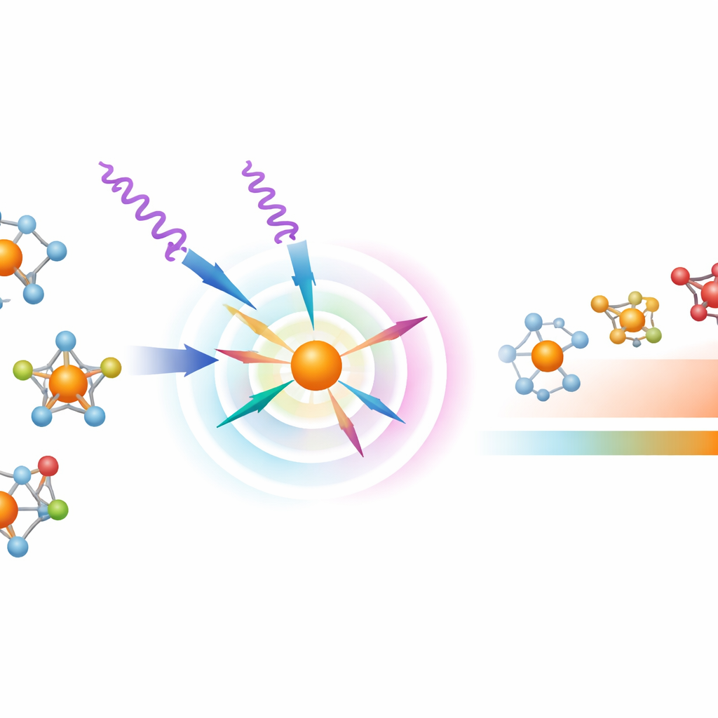
What this means for future radiopharmaceuticals
In accessible terms, this work demonstrates that carefully crafted X-ray measurements can tell not just where atoms sit around a metal ion, but how tightly and cooperatively they share electrons. For radiopharmaceuticals, that sharing governs how firmly a radioactive atom is held inside its molecular cage as it travels through the bloodstream and into tumors. The framework developed here—using lanthanum stand-ins, high-resolution spectroscopy, and matching theory—provides a roadmap for judging and improving new chelators before scarce actinium is ever introduced. While the electronic structure of actinium itself is more complex, the same style of measurements at different X-ray edges should help disentangle its bonding behavior. Ultimately, such insights are expected to guide the design of next-generation cancer therapies that are both more potent against tumors and safer for patients.
Citation: Ramanantoanina, H., Schacherl, B., Kovács, A. et al. High-energy resolution X-ray spectroscopy reveals bonding characteristics of La3+ homologues of actinium radiopharmaceuticals. Commun Chem 9, 148 (2026). https://doi.org/10.1038/s42004-026-01929-4
Keywords: actinium radiopharmaceuticals, lanthanum complexes, X-ray spectroscopy, metal–ligand bonding, targeted alpha therapy