Clear Sky Science · en
Molecular mechanisms and multi-organ regulatory roles of anti-Müllerian hormone in female reproduction
Why this hidden hormone matters
Anti-Müllerian hormone, or AMH, is best known today as a blood test that hints at how many eggs a woman has left. This review argues that AMH is far more than a simple fertility number. It acts as a flexible control signal that links the ovaries, brain, uterus, and placenta, helping to coordinate when eggs wake up, how hormones pulse, and how the womb prepares for pregnancy. Understanding this wider role may change how we interpret AMH tests and how we think about conditions such as polycystic ovary syndrome and age-related fertility decline.
A traffic controller for reproductive signals
AMH is made mainly by cells that surround immature eggs in the ovary. For years, it was viewed as a local brake that slows the early growth of follicles so the egg supply is not used up too quickly. The authors propose a broader view: AMH behaves as a “signaling hub” whose effects depend on where and when it is acting. After AMH is produced, it circulates in a protected form that can travel in the bloodstream while also building up in the fluid inside follicles. By binding to a dedicated receptor on cell surfaces, it launches a chain of events inside the cell that turns specific genes on or off. Built-in feedback molecules keep this signaling in check so it does not become too strong or too weak.
How AMH talks to other cellular switches
Inside cells, AMH mainly works through a family of proteins called Smads that relay messages from the cell surface to the DNA in the nucleus. But AMH does not act alone. It interacts with other major pathways, including Wnt/β-catenin and MAPK, which are involved in cell growth, survival, and hormone production. In some settings, this crosstalk helps reshape tissues, as in the regression of embryonic ducts in males. In the ovary, it can slow cell division and push damaged cells toward self-destruction, effects that may be useful for limiting tumor growth. AMH signaling is also shaped by the body’s internal clock, metabolic state, and inflammation, hinting that nutrition, body fat, and immune signals can all influence how AMH behaves.
Hormones, metabolism, and vitamin D as fine-tuners
AMH levels are tightly woven into the broader hormone web. Follicle-stimulating hormone (FSH) helps follicles grow but, once it rises beyond a threshold, it lowers AMH output from the same cells it is stimulating. Estrogen further suppresses AMH, easing the way for follicles to mature. Transcription factors such as SF1 and FOXL2 bind directly to the AMH gene switch and work together to set its baseline activity, while other factors control the AMH receptor. Metabolic hormones produced by fat tissue, including leptin and adiponectin, and inflammatory molecules such as TNF-α and interleukin-6 also adjust AMH production and action, especially in conditions like polycystic ovary syndrome. Vitamin D adds another layer: it can bind to specific sites on the AMH gene and appears to raise AMH levels in some contexts, while also changing how AMH signals inside ovarian cells.
From brain signals to egg survival and pregnancy
AMH shapes reproduction at every level, from brain circuits to local tissue environments 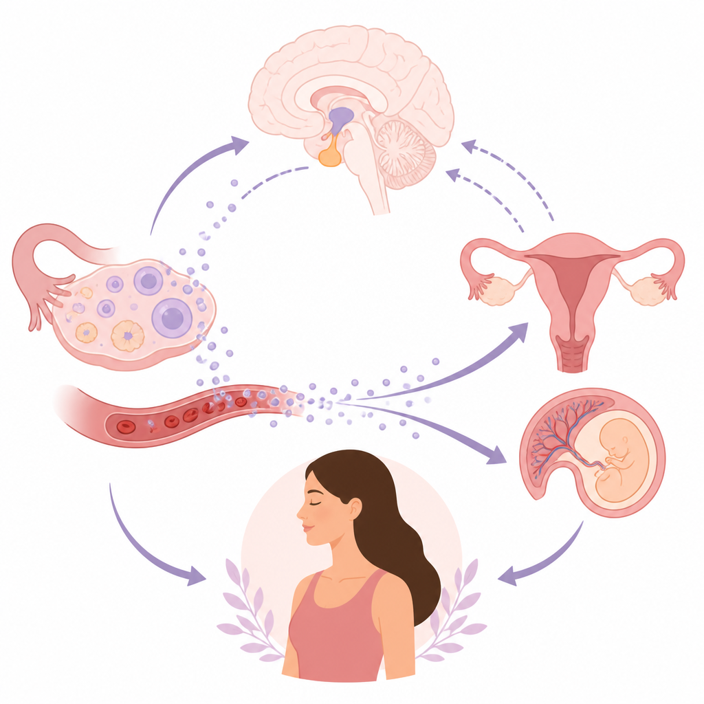
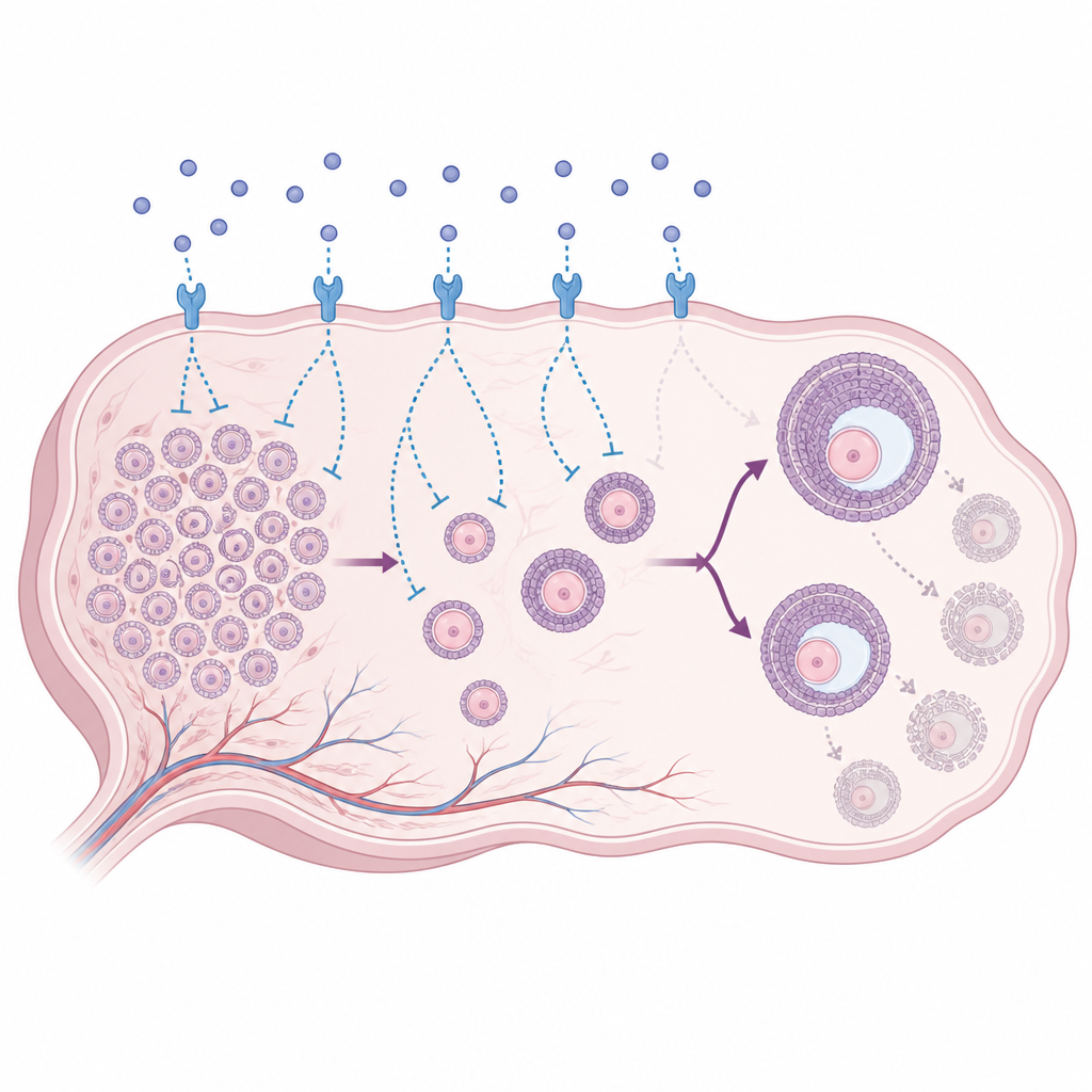
Rethinking a familiar fertility test
The authors conclude that AMH should no longer be seen only as a tally of remaining eggs. Instead, it is a context-dependent coordinator that links the size of the ovarian reserve to brain hormone rhythms, ovarian responsiveness, and the readiness of the uterus and placenta. Because AMH levels are shaped by genetics, assay methods, hormones, metabolism, inflammation, and vitamin D status, a single blood value cannot be interpreted in isolation. Future research that targets AMH or its receptor in specific tissues, combined with detailed molecular analyses, may open new ways to understand and possibly manage disorders such as polycystic ovary syndrome, premature ovarian insufficiency, and fertility changes with age.
Citation: Li, J., Zhu, W., Bu, Y. et al. Molecular mechanisms and multi-organ regulatory roles of anti-Müllerian hormone in female reproduction. Commun Biol 9, 658 (2026). https://doi.org/10.1038/s42003-026-10273-1
Keywords: anti-Müllerian hormone, ovarian reserve, female reproduction, polycystic ovary syndrome, hypothalamic pituitary axis