Clear Sky Science · en
Insights into unique anatomical structures of the ascidian Halocynthia papillosa obtained by multimodal imaging
Sea squirts as living windows into our own past
On rocky Mediterranean reefs, the bright red sea squirt Halocynthia papillosa looks like a simple, motionless blob. Yet this unassuming animal is one of our closest invertebrate relatives, sitting on the evolutionary branch just beside vertebrates. Understanding how its body is built can reveal how early chordates were put together and how today’s marine animals cope with changing oceans. This study uses a suite of modern imaging methods, from MRI scanners to powerful X‑ray microscopes, to uncover hidden structures in the sea squirt’s armor, nerves, and feeding tentacles.
Peering inside a reef-dwelling filter
Rather than relying on thin tissue slices alone, the researchers combined multiple ways of looking at the same animals. Conventional light microscopes and a technique called Thunder microscopy provided sharp two-dimensional overviews of whole sea squirts and their tissues. Magnetic resonance imaging (MRI), similar to scans used in hospitals, produced three-dimensional views of the entire animal, clearly separating the tough outer coat from the soft inner body. A synchrotron-based method called high-throughput tomography (HiTT) added extremely fine X-ray detail, while confocal microscopy captured natural glow from certain tissues without any added dyes. Together, these approaches let the team zoom from the scale of the whole animal down to structures only a few micrometers across. 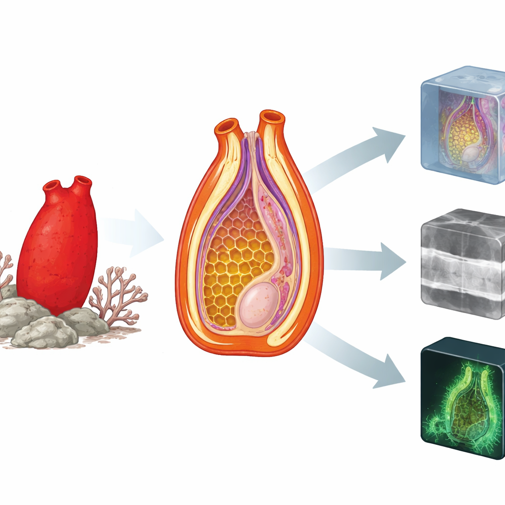
An unexpected armor of glowing spines
The outer coat, or tunic, of Halocynthia papillosa turned out to be much more intricate than a simple skin. Inside this coat, the scientists saw layered sheets of cellulose, the same basic material found in plant cell walls. Near the surface, the layers twist into spiral depressions that support conical spines, forming a kind of three-dimensional scaffold. These spines are crowned by a cuticular layer that, under blue-green light, glows strongly on its own. In relaxed animals, the glowing patches are separated by dark gaps, but when the animals contract, the spines shift and overlap, creating a nearly continuous fluorescent shield over the surface. Spectral measurements showed that contracted animals reflect much more light, especially on the more strongly colored side of the body, suggesting that muscle-driven changes in shape and pigment exposure may alter how the animal appears against the reef background.
Hidden nerve cords and a puzzling brain
Inside, the team focused on the central nerve cord that links the two siphons through which water flows. In many related sea squirts, this cord swells into a distinct brain-like knot called the cerebral ganglion. In Halocynthia papillosa, however, even high-resolution X-ray scans did not reveal any such obvious thickening; instead, a long, uniform cord runs between two branching points near the siphons. This cord splits repeatedly and then wraps around each siphon as a ring, with muscle fibers arranged in ordered bundles alongside it. A structure called the dorsal tubercle, perched just in front of the oral siphon, forms a jelly-like funnel with raised horns and sits directly above one of these branching points. Earlier work in other species suggests this region likely houses the main concentration of nerve cells, but here it cannot be distinguished by shape alone, hinting at a different organization of the “brain” in this species.
Feathery tentacles that feel and feed
Around the mouth opening, the researchers reconstructed the sea squirt’s oral tentacles in three dimensions. These finger-like structures form a ring facing the incoming water stream and bear smaller side branches on their underside. The tentacles are rounded toward the outer side, where water enters, and become flatter toward the inside of the body, a shape that likely steers flow while forming a continuous sensory fringe. Within each tentacle, HiTT imaging revealed a paired system of larger tubes: one for blood and one for nerves. The blood vessels branch neatly into each side branch of the tentacle, while a matching nerve pattern runs along the opposite side. This layout supports the idea that Halocynthia papillosa has a largely closed or semi-closed circulation and that its tentacles are both filters and sensitive detectors of what passes through the mouth.
Why these details matter for reefs and for us
By combining several cutting-edge imaging tools, this study paints a detailed picture of how a common Mediterranean sea squirt is put together, from its spiralized, glowing armor to its unusual central nerve cord and finely wired feeding tentacles. These anatomical twists show that even among closely related sea squirts, there is more diversity than the few standard laboratory species suggest. Because ascidians help move nutrients through reef ecosystems and are used as indicators of pollution, warming, and noise, understanding their real anatomical variety matters for both ecology and environmental monitoring. At the same time, as one of our nearest invertebrate cousins, Halocynthia papillosa offers a fresh window into how early chordate bodies—and their nervous systems and protective coats—may have evolved. 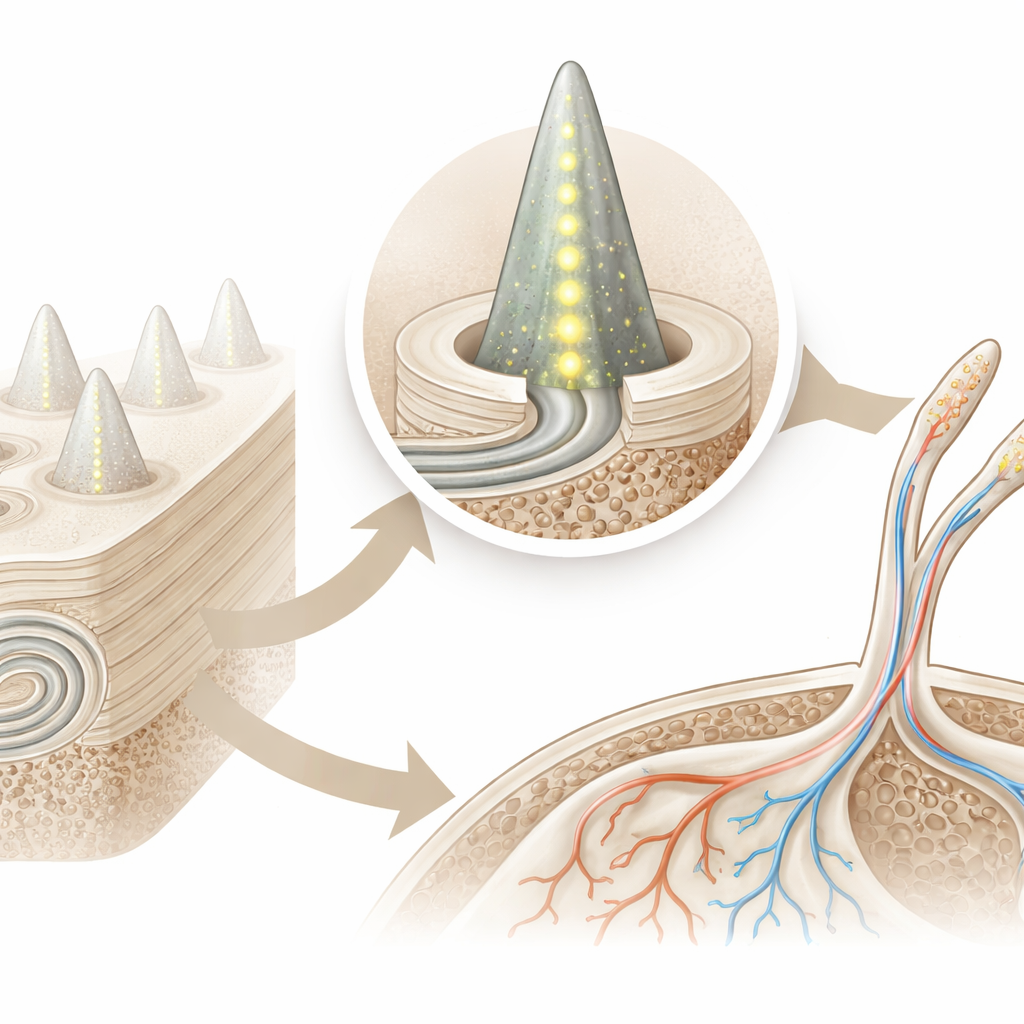
Citation: Hessel, L., Albers, J., Michalek, A. et al. Insights into unique anatomical structures of the ascidian Halocynthia papillosa obtained by multimodal imaging. Commun Biol 9, 557 (2026). https://doi.org/10.1038/s42003-026-10102-5
Keywords: ascidian anatomy, marine imaging, sea squirt tunic, nervous system, reef ecology