Clear Sky Science · en
Structural basis of substrate recognition and membrane association by the bacterial lysyl-phosphatidylglycerol hydrolase AcvB
How Bacteria Tune Their Skin
Just like humans put on a raincoat or sunscreen, bacteria adjust their outer "skin" to survive in harsh surroundings. This study looks at how a plant-infecting bacterium fine-tunes the oily molecules in its membrane so it can withstand acid and natural antibiotics. By uncovering the three-dimensional shape of a key enzyme, the research reveals how bacteria keep their protective coating in balance and hints at new ways to disarm harmful strains that attack crops.
Why Bacterial Coats Matter
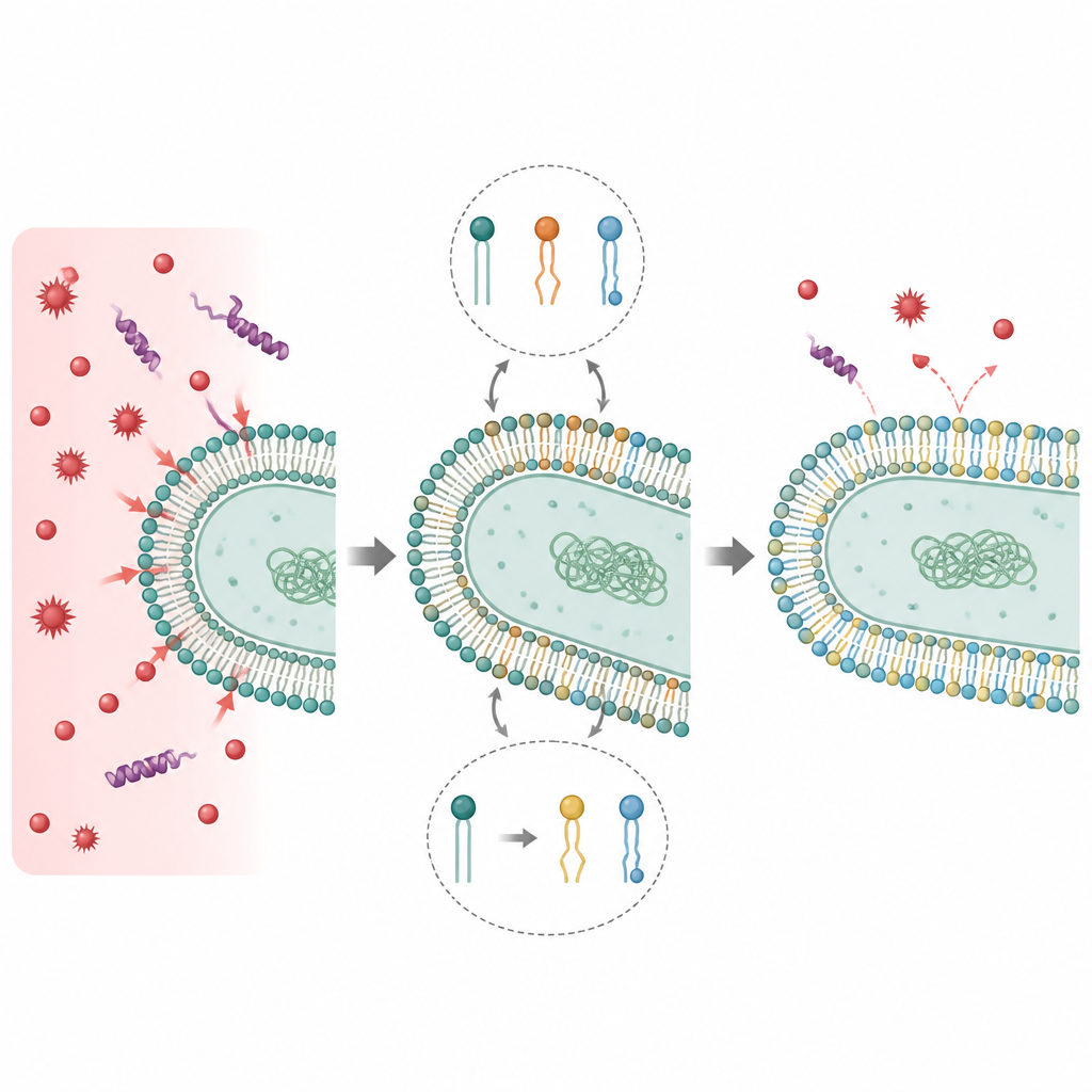
Bacterial membranes are built from a mix of oily molecules that not only hold the cell together but also help it adapt to threats such as temperature changes and hostile chemicals. Many bacteria protect themselves from positively charged antimicrobial peptides, natural defense molecules made by plants and animals, by decorating one common membrane fat, phosphatidylglycerol, with amino acids like lysine. These modified fats, especially lysyl-phosphatidylglycerol, reduce the overall negative charge of the membrane surface, making it harder for antimicrobial peptides to latch on and punch holes in the cell.
Balancing Protection and Growth
In the plant pathogen Agrobacterium tumefaciens, one protein called LpiA adds lysine to membrane fats, while another protein, AcvB, removes it. Together they act as a molecular dial that sets how much lysyl-phosphatidylglycerol the membrane contains. If AcvB is missing, earlier work showed that the bacterium accumulates too much of the modified lipid, grows poorly in acidic conditions, and loses its ability to cause plant tumors. This makes AcvB essential for keeping the membrane in a sweet spot where the cell is protected from stress but still able to carry out key tasks like transferring DNA into plant cells.
Seeing the Shape of a Membrane Helper
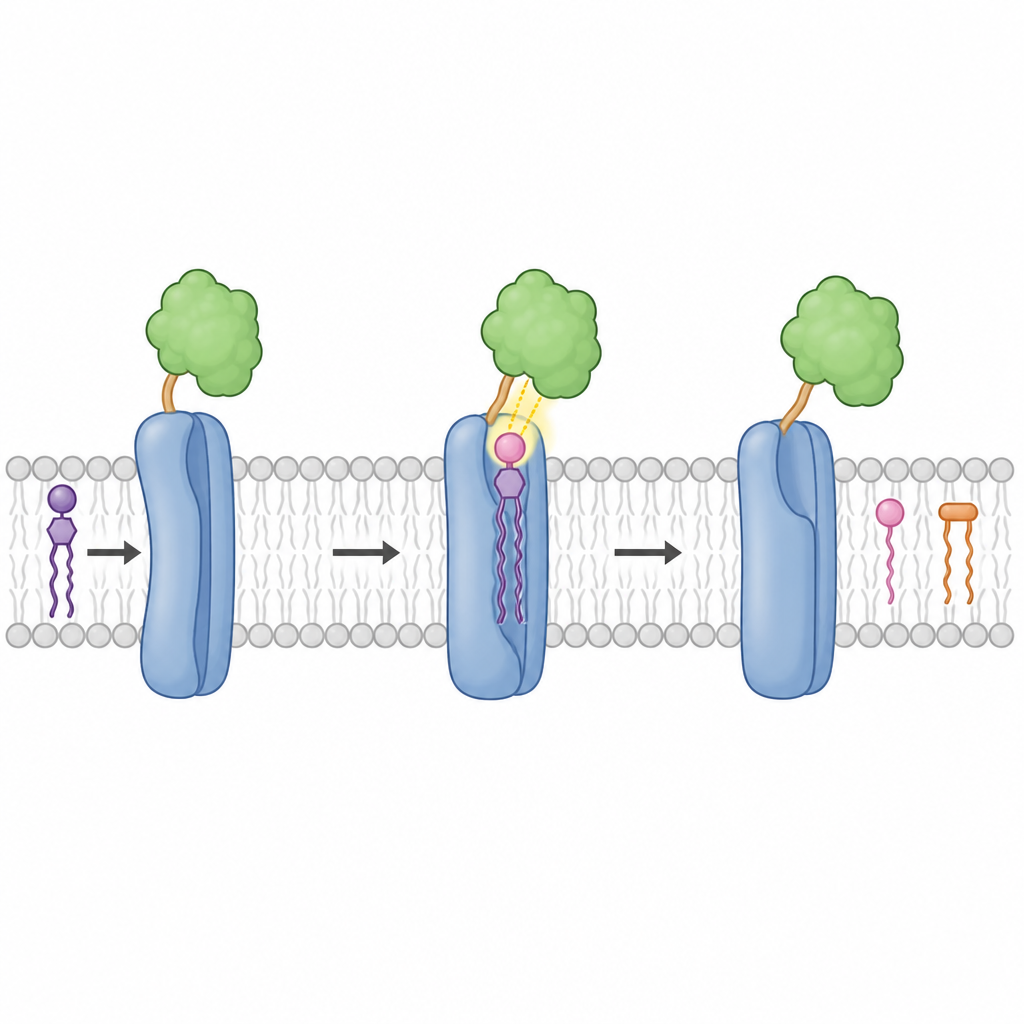
To understand how AcvB works, the researchers determined high-resolution crystal structures of the mature enzyme and of its active portion. AcvB turned out to have two similar lobes, with the back half forming the catalytic heart of the protein. This catalytic region contains a negatively charged cavity that cradles the positively charged head of lysyl-phosphatidylglycerol. Within this pocket, a handful of acidic amino acids precisely position the lysine group so that a pair of catalytic residues can attack and break the chemical bond that links lysine to the fat molecule, releasing free lysine and restoring the original membrane lipid.
How AcvB Grabs the Membrane
Although AcvB floats in the watery space between the bacterial inner membrane and the cell wall, its target lipid is embedded in the membrane itself. The crystal structure revealed a short loop near the active site that sticks out from the protein surface and carries two oily amino acids, tryptophan and leucine. Experiments showed that this loop enables the enzyme to anchor briefly in the membrane, bringing the active site close enough to reach its buried substrate. When the loop was deleted, or when its oily side chains were replaced with more water-loving ones, AcvB no longer associated well with membranes and could not efficiently remove lysine from the lipid.
Teamwork Between Two Enzymes
The study also found that AcvB physically interacts with LpiA, the enzyme that adds lysine to the same lipid. Tests with purified proteins indicated that the active back half of AcvB makes the main contact with LpiA. This partnership does not depend on LpiA's own chemistry, suggesting that LpiA serves partly as a docking site that tethers AcvB to the membrane. By placing the remover enzyme next to the adder enzyme, the cell can quickly raise or lower the amount of lysyl-phosphatidylglycerol in response to changes such as drops in pH or exposure to antimicrobial peptides, without overcommitting to either extreme.
What This Means for Plant Health
Altogether, the work reveals how the detailed shape of AcvB allows it to recognize a charged lipid head, touch the membrane just enough to work, and cooperate with its partner LpiA. For non-specialists, the key message is that bacteria use finely tuned molecular switches on their surface to balance defense and growth. Understanding this balancing act at the atomic level provides a roadmap for designing compounds that disrupt it, which could weaken plant-pathogenic bacteria by preventing them from adjusting their protective coats.
Citation: Hoshi, M., Matsumoto, D. & Watanabe, Y. Structural basis of substrate recognition and membrane association by the bacterial lysyl-phosphatidylglycerol hydrolase AcvB. Commun Biol 9, 689 (2026). https://doi.org/10.1038/s42003-026-10087-1
Keywords: bacterial membrane, lysyl phosphatidylglycerol, AcvB enzyme, LpiA interaction, antimicrobial resistance