Clear Sky Science · en
MIMIC: a flexible pipeline to register and summarize IMC-MSI experiments
Seeing More Inside Tissues
Modern microscopes can show us where cells sit in a tissue, while chemical imaging can reveal which molecules are present. Until now, it has been hard to merge these views into a single, reliable picture. This paper introduces a new workflow called MIMIC that helps scientists precisely line up and combine these different kinds of images so they can better understand how cells and molecules interact in health and disease.
Why Combining Views Matters
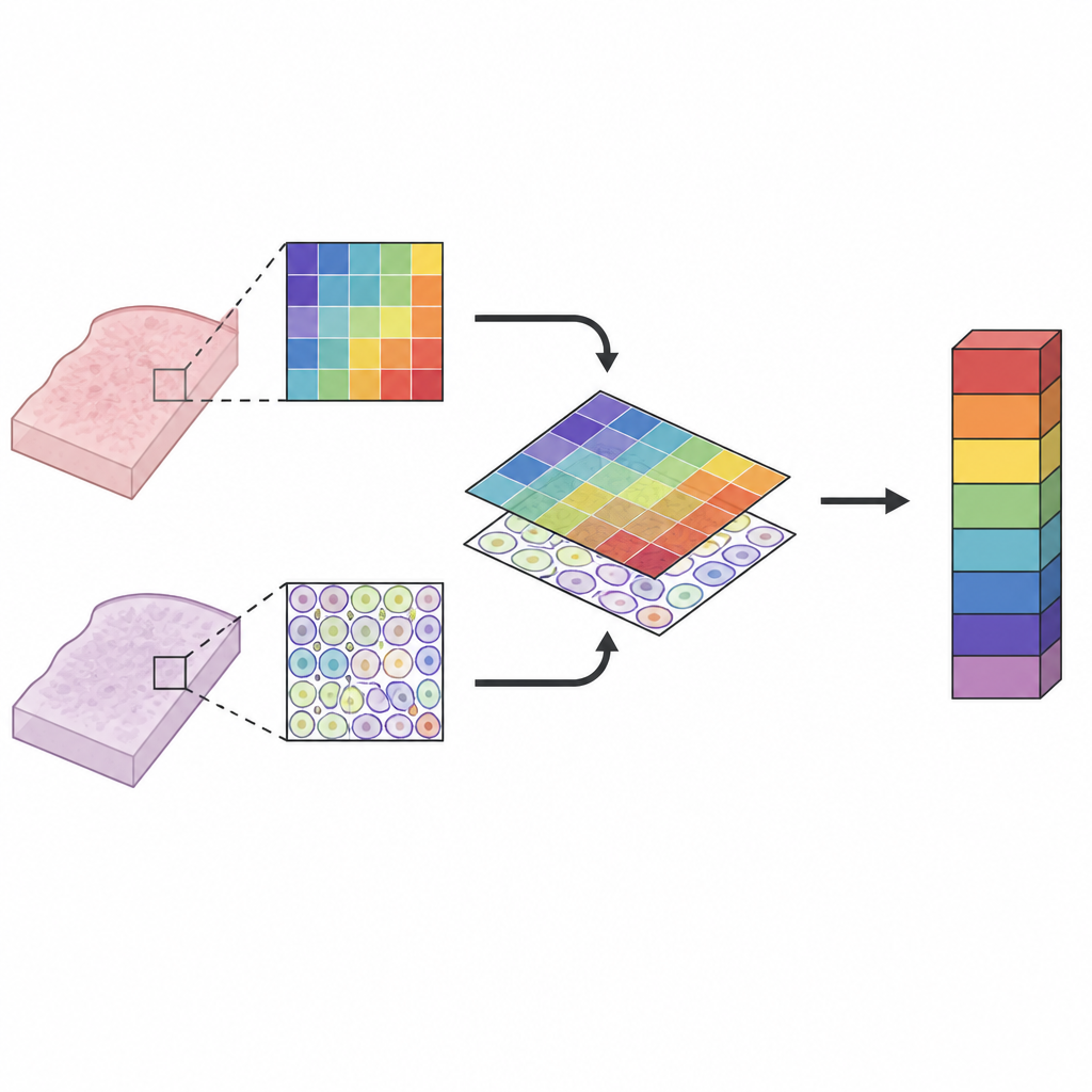
Biologists want to know not just which cells and molecules are in a tissue, but where they are and how they share space. One set of tools, such as imaging mass cytometry, can identify many cell types by their proteins at near single cell detail. Another, mass spectrometry imaging, maps a wide range of molecules like fats and sugars across the tissue. Each method has strengths and weaknesses in speed, sensitivity, and sharpness. Used alone, each gives only part of the story. Used together, they can reveal how cell neighborhoods and local chemistry shape disease, but only if their images can be aligned with high precision.
A Careful Step by Step Alignment
MIMIC offers a semi-automated pipeline to bring these images into the same frame. The authors use regular light microscope scans taken before and after each measurement as a scaffold. First, they detect the tiny burn marks left by the mass spectrometry laser and match them to the coarse chemical pixel grid. Then they register the brightfield scans taken before and after both imaging steps, using a series of geometric transformations that shift, rotate, and smoothly warp the images as needed. Finally, they place the high detail cell map from imaging mass cytometry onto the matching brightfield image. Chaining these transformations lets them map each chemical pixel onto local cell types across the tissue.
Checking That the Match Is Tight
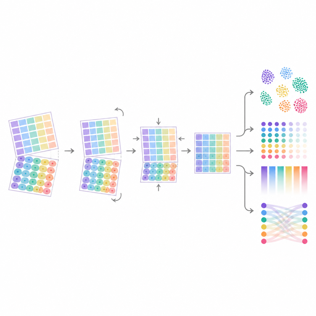
Because later analyses depend on this alignment, MIMIC spends a lot of effort measuring how accurate it is. The team compares known landmarks, such as matching features or cell nuclei, between image pairs and calculates how many micrometers they are apart after registration. Most steps, such as linking laser marks to pixels or matching images taken before and after one experiment, reached median errors of only about two micrometers, close to the size of a single cell nucleus. More difficult steps, like matching neighboring tissue slices, were less precise and sometimes required manual review. The authors also show that fully automatic methods often outperform simple manual alignment based on only a few points, especially when working with low resolution scans.
From Pixels to Cell Molecule Links
Once images are aligned, MIMIC moves from geometry to statistics. For every chemical signal and every tissue pixel, the workflow notes which cell types share that pixel, then fits spatial models that account for the fact that nearby pixels tend to be similar. This first step estimates how strongly each molecule is associated with each cell type on a given slide. A second modeling step then compares these association strengths across many samples and conditions. Simulations show that as registration errors grow, these estimated links weaken and become less reliable, underscoring the need for the strict quality control that MIMIC provides.
Proof in Artificial and Diseased Tissues
The authors test MIMIC in three settings. In a synthetic “tissue” made from three known cell lines, the pipeline recovers expected matches between specific lipids and each cell line, even when the cells are mixed together. Reanalyzing a public dataset, they show that improved automatic alignment leads to more consistent chemical signals per cell and slightly stronger statistical evidence for cell molecule associations. Finally, they apply MIMIC to human liver samples from patients with advanced metabolic liver disease. Here, the method rediscovers known patterns: certain sugar-coated molecules concentrate in regions rich in liver cells, while others are tied to immune cell areas. It also highlights additional, more subtle links that only become visible when working at the pixel level instead of averaging over large tissue zones.
What This Means for Future Studies
In plain terms, MIMIC is a set of tools and checks that let scientists confidently overlay “who is where” with “what molecules are where” inside tissues. By tightening up the image alignment and offering a clear route from raw pictures to statistical summaries, it makes it easier to discover how specific cell types and local chemistries go together. This can deepen our understanding of complex diseases such as liver disorders and can be extended to other spatial methods. MIMIC does not cure disease by itself, but it provides a sturdy mapmaking kit for researchers exploring the cellular and molecular landscape of tissues.
Citation: Gerber, R., Griner, J., Guglietta, S. et al. MIMIC: a flexible pipeline to register and summarize IMC-MSI experiments. Commun Biol 9, 712 (2026). https://doi.org/10.1038/s42003-026-09961-9
Keywords: spatial omics, image registration, mass spectrometry imaging, imaging mass cytometry, liver tissue