Clear Sky Science · en
Scalable discovery of spatial multicellular patterns via neighborhood-to-sequence transformation
Finding hidden order in crowded tissues
Modern microscopes can now capture the exact position and molecular makeup of millions of cells in a tissue slice. But spotting the important recurring arrangements of cells by eye is nearly impossible. This study introduces a new way to scan these massive maps and automatically uncover the small, repeated “neighborhoods” of cells that distinguish healthy tissue from disease, recovery from aging, or one organ zone from another.
From cell neighborhoods to simple sequences
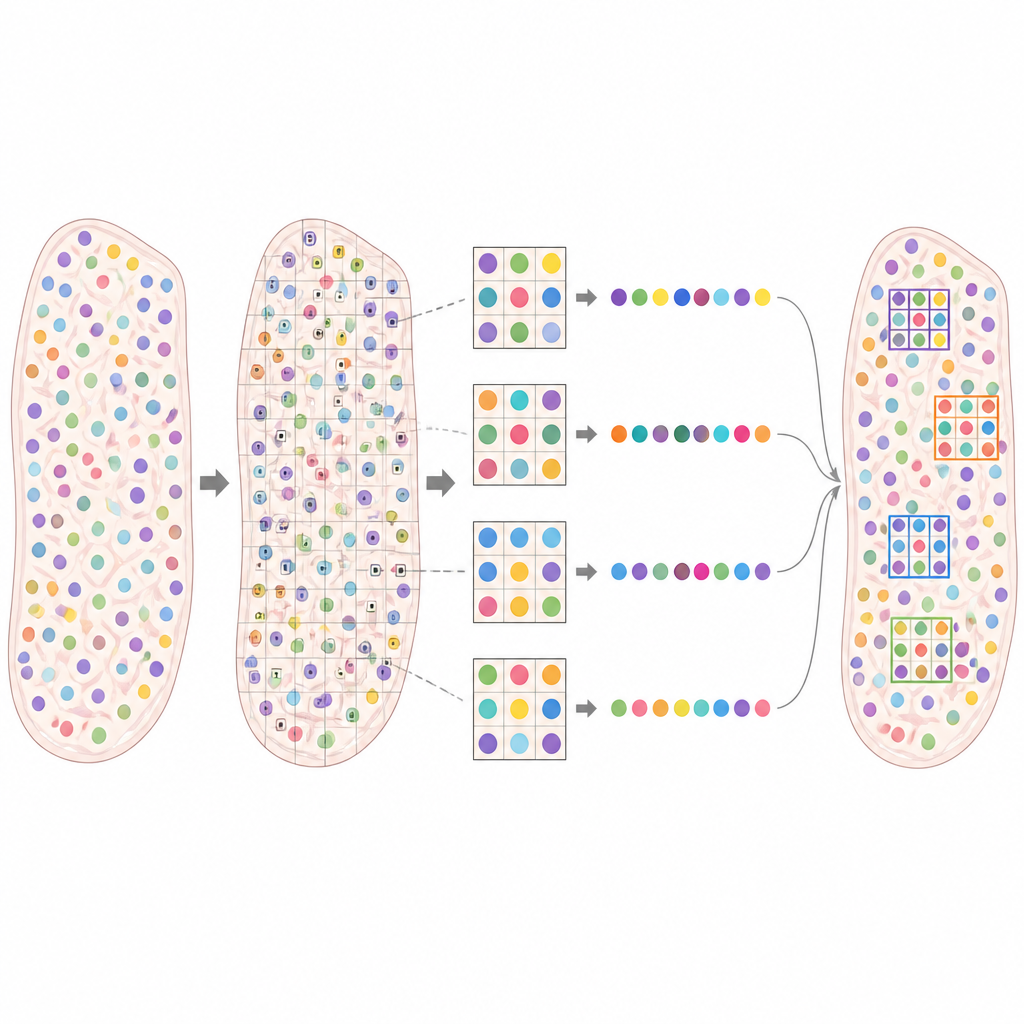
The authors present a computational framework called FDPMining that turns the problem of reading complex tissues into a more manageable data mining task. Instead of looking at cells one by one, the method builds a small grid around every cell, recording which cell types are found in each surrounding tile. A clever “Neighborhood to Sequence” strategy then converts each grid into a unique list of numbers that still fully preserves which cell types sit next to which, and where. Because this transformation is lossless and reversible, the computer can search these lists for recurring combinations and then map any interesting pattern back into its original position in the tissue.
Hunting for frequent and distinctive patterns
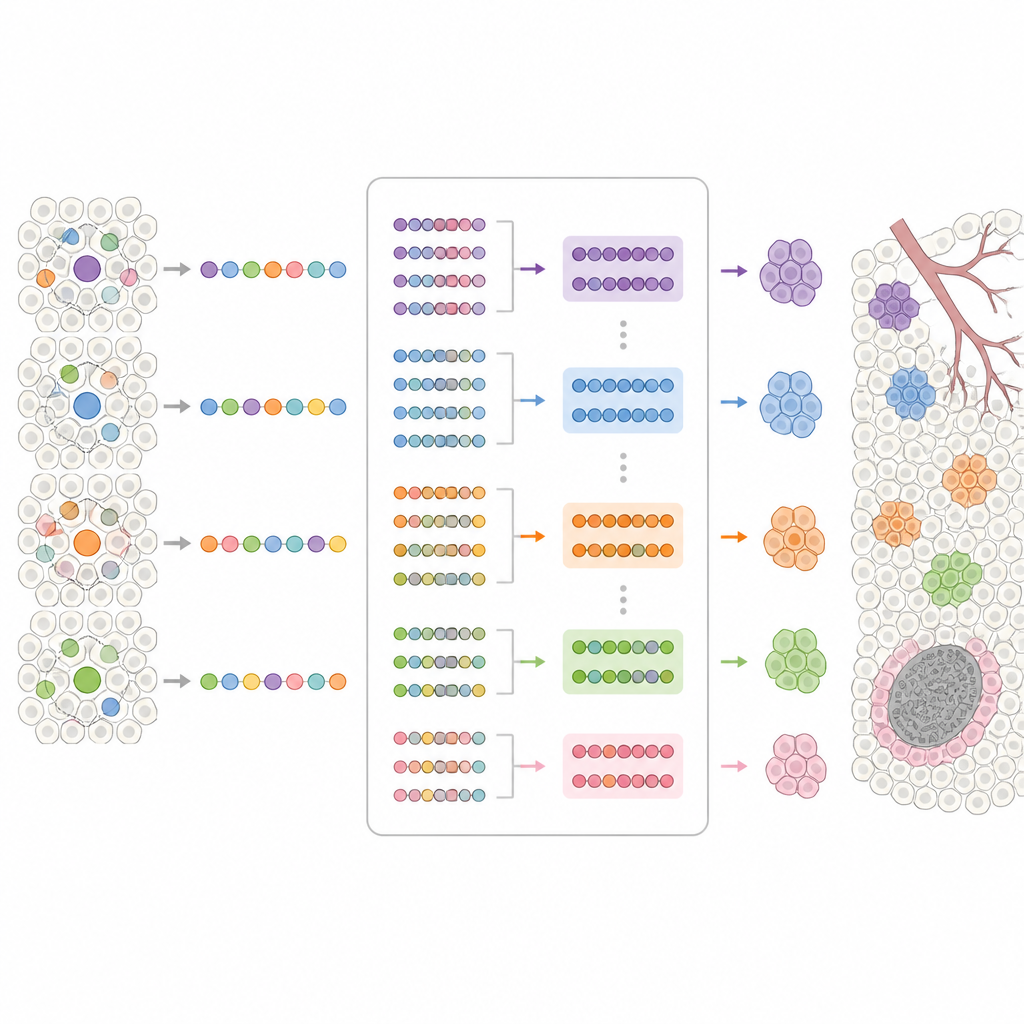
Once every neighborhood has been encoded, FDPMining searches for multicellular arrangements that are both common and strongly tied to a particular condition, such as a disease state or a specific anatomical region. The framework borrows tools from market basket analysis, where algorithms learn which products are often bought together. Here, “items” are cell types at particular locations in the grid, and “transactions” are all the neighborhoods in a tissue. The method identifies patterns that appear repeatedly but mainly in one condition and rarely in others. These “frequent and distinctive patterns” can then be projected back onto the tissue slice, making it easy for researchers to see where they occur and what larger structures they might belong to.
Testing the approach across many tissues
The team showed that FDPMining works across a wide range of spatial omics technologies and tissues, from human brain to mouse embryo. In simulated data, the method picked up subtle rearrangements of cell layers that other tools missed, proving that it can recognize fine shifts in local tissue architecture. In axolotl brain, it revealed changing partnerships between wound-activated neurons and different neuron subtypes as regeneration progressed, and it showed how specific oligodendrocyte subtypes reorganize with age in the mouse brain. In colorectal cancer samples, FDPMining separated patients with better outcomes, whose tumors contained repeated neighborhoods rich in B cells and helper T cells, from patients whose tumors were dominated by neighborhoods of myeloid cells linked to a more suppressive immune environment.
Zooming in on landmarks like vessels and plaques
The authors also used FDPMining to focus on key landmarks inside tissues. In mouse liver, they labeled neighborhoods by their proximity to central or portal veins and found that distinct hepatocyte subtypes and supporting cells formed characteristic patterns around each vessel type, echoing the well-known concept of liver zonation. In a mouse model of Alzheimer’s disease, they labeled neighborhoods by whether they lay near amyloid plaques and uncovered repeated combinations of microglia, excitatory neurons, and other glial cells around plaques. These plaque-centered patterns matched known “core and shell” arrangements and were largely absent in healthy tissue, underlining their tight link to disease microenvironments.
Why these hidden patterns matter
By systematically scanning tissues for repeated cell neighborhoods that are tied to specific conditions or landmarks, FDPMining offers a powerful lens on how cells cooperate in health and disease. Rather than only outlining broad tissue zones, it pinpoints the small multicellular building blocks that make up those zones and change in response to injury, aging, cancer, or neurodegeneration. For non-specialists, the message is that there is a discoverable “grammar” to how cells arrange themselves, and this method helps read that grammar at scale, opening paths to new spatial biomarkers and richer biological insight.
Citation: Zhao, F., Wang, S., Wang, Z. et al. Scalable discovery of spatial multicellular patterns via neighborhood-to-sequence transformation. Commun Biol 9, 725 (2026). https://doi.org/10.1038/s42003-026-09923-1
Keywords: spatial omics, cell neighborhoods, tissue microenvironment, pattern mining, Alzheimer’s disease