Clear Sky Science · en
Expanded iOn switch toolkit enables flexible clonal labeling and dynamic imaging in model and non-model animals
Following Family Trees in the Growing Brain
How does a single immature brain cell give rise to the incredible variety of neurons that wire up our thoughts, senses, and memories? To answer this, scientists need ways to mark the “family trees” of cells as the brain develops, not only in classic lab species like mice but also in animals that better reflect natural diversity. This study introduces an upgraded toolkit that lets researchers color and track brain cell lineages with unusual flexibility across many different vertebrates.
A Color-Coded Map of Cell Families
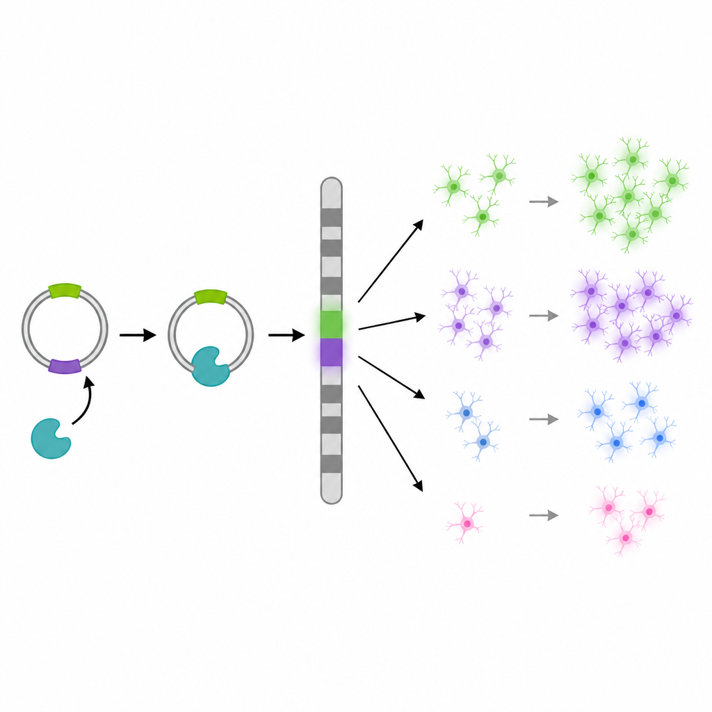
At the heart of the work is an improved version of the “iOn switch” system, a DNA-based tool that turns on fluorescent markers only when they are stably built into a cell’s genome. This is important because it filters out short-lived signals that fade as DNA pieces are lost, leaving only cells that have truly taken up the label. The authors redesigned and tuned this system so it can be used to follow clones—groups of cells descended from a single progenitor—in developing brains, an approach that reveals how basic building blocks of brain architecture are assembled.
Dialing Labels from Sparse to Dense
A key advance is that the same toolkit can now be used for both very sparse and very dense labeling, simply by changing how much DNA is delivered. In chick and mouse brains, high DNA doses produced dense color patterns that blanket large areas, useful for reconstructing many overlapping clones at once. Lower doses yielded only a few scattered labeled clusters, ideal for isolating single family trees without confusion. Tests in cultured cells helped the team identify DNA and enzyme ratios that keep labeling efficient while avoiding damage to cells, showing that the switch can be both powerful and gentle.
Adding More Colors and Cell Landmarks
The researchers also broadened the color palette. In addition to red, the system now supports cyan-like and yellow fluorescent proteins, and keeps an infrared channel free for future use. By combining these colors, individual clones can be recognized by unique mixes of hues. On top of this, the team created variants that target different parts of the cell, such as the nucleus, cell membrane, or mitochondria. This allows scientists to see both who is related to whom and where exactly within each cell the fluorescence appears, making it easier for software to separate and measure neighboring cells in crowded brain tissue.
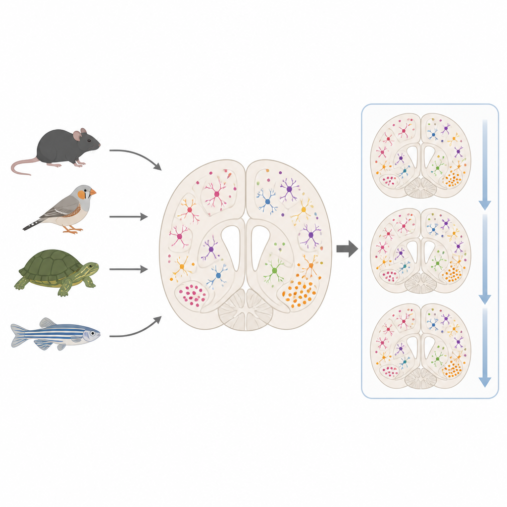
Reaching Beyond Classic Lab Animals
To show that the toolkit is not limited to a single species, the team tested it in a broad panel of vertebrates. Using electrical pulses or microinjection to deliver the DNA, they obtained clear multicolor labeling in chicks, turtles, rats, guinea pigs, mice, and zebrafish. In each case, labeled neurons and glial cells could be distinguished by their shapes, and the colors remained stable long enough for detailed imaging. The system also provided smoother, more uniform signals than traditional plasmid methods during time-lapse movies, allowing researchers to track migrating brain cells over days without constantly adjusting microscope settings.
What This Means for Understanding Brain Diversity
In simple terms, this updated iOn switch toolkit is a flexible set of genetic “highlighters” that can mark, distinguish, and follow brain cell families across a wide range of animals. By tuning label density, mixing colors, and targeting specific cell compartments, researchers can now design lineage experiments that fit both simple and complex questions, from single-family tracking to dense reconstructions of whole regions. Because the same approach works in model and non-model species, it opens the door to side-by-side comparisons of how different brains grow and evolve, helping us understand how varied neural structures arise from similar developmental rules.
Citation: Ngiam, Z.C., Wada, K., Hatakeyama, J. et al. Expanded iOn switch toolkit enables flexible clonal labeling and dynamic imaging in model and non-model animals. Commun Biol 9, 654 (2026). https://doi.org/10.1038/s42003-026-09907-1
Keywords: lineage tracing, brain development, fluorescent labeling, evo devo, neural progenitors