Clear Sky Science · en
Ontogeny independent expression of LPCAT2 in granuloma macrophages during experimental visceral leishmaniasis
Why liver inflammation in infection matters
When certain infections linger in the body, our immune system walls them off in small structures called granulomas. In visceral leishmaniasis, a serious tropical disease caused by the parasite Leishmania donovani, these structures form in the liver and can help keep the infection under control. Understanding how cells inside granulomas use energy and change their behavior could open doors to better ways to manage inflammation without completely shutting down our defenses.
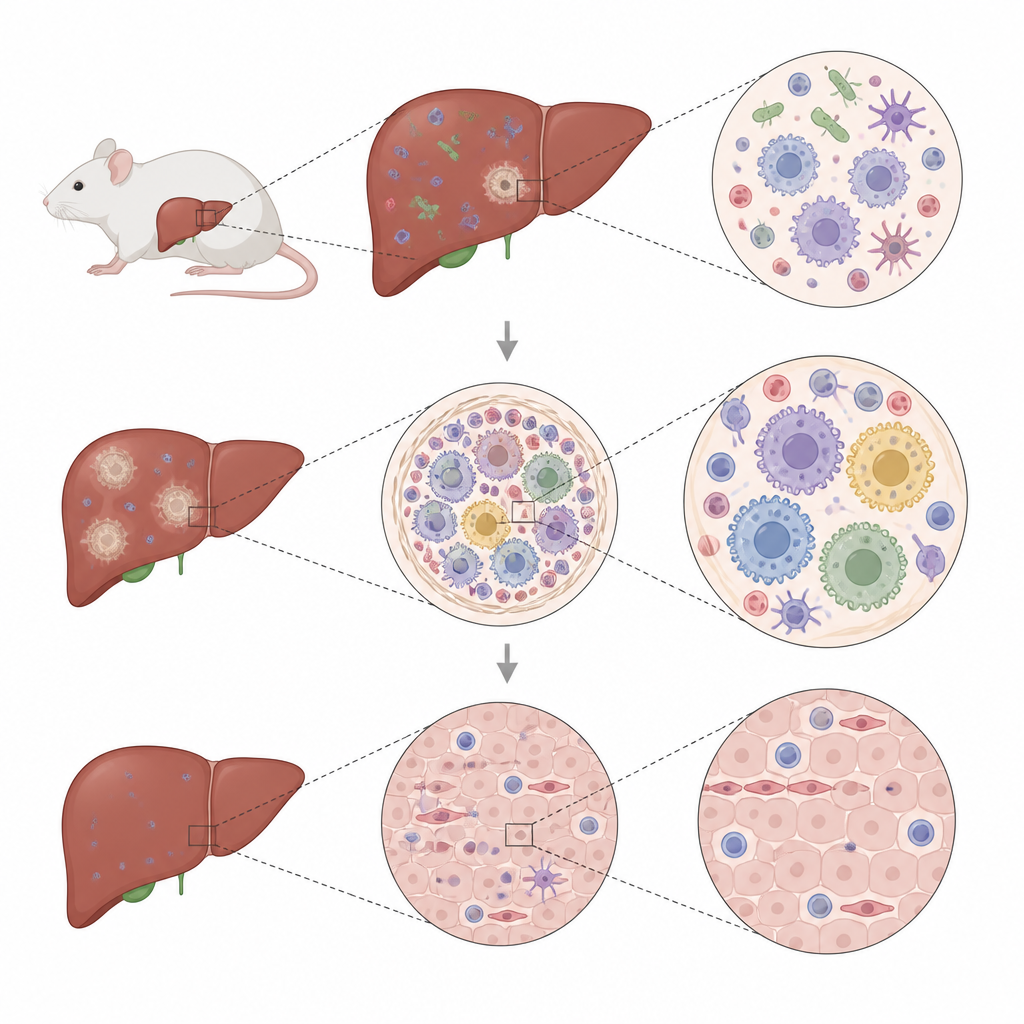
Tiny immune fortresses inside the liver
The authors used a well established mouse model of visceral leishmaniasis, where parasites settle in liver cells called Kupffer cells and trigger the formation of organized granulomas. These structures are crowded neighborhoods of immune cells, including resident liver macrophages, incoming monocyte derived macrophages, T cells, and a few neutrophils. Granulomas help limit parasite growth, but their success depends on how these cells switch between attack and repair modes over time, a process that places heavy demands on how they handle fuel and building blocks inside the tissue.
Looking at genes, fats, and proteins all at once
To map what happens inside and around granulomas, the team combined several advanced imaging methods. They overlaid spatial gene activity maps with mass spectrometry images that show the distribution of many lipids, as well as single cell gene profiles and protein measurements from sorted macrophages. By aligning these layers at nearly the same locations in liver slices, they could see which lipids appeared where particular cell types and gene programs were active. This multimodal view highlighted specific spots that matched the histological outline of granulomas and revealed that both gene expression and lipid patterns were clearly organized in space.
A membrane shaping enzyme in the spotlight
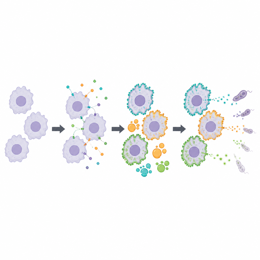
Within granulomas, the study focused on an enzyme called LPCAT2, which helps remodel the phospholipids that make up cell membranes. Both resident Kupffer cells and incoming monocyte derived macrophages showed high levels of the gene for LPCAT2 and related enzymes that process a group of lipids known as lysophosphatidylcholines. Mass spectrometry imaging showed that these lipids and their products accumulated particularly in granuloma regions. Microscopy confirmed that LPCAT2 protein was concentrated in granuloma macrophages, regardless of whether they had been in the liver since development or had recently arrived from the blood.
Linking membrane changes to inflammatory behavior
Macrophages with strong LPCAT2 activity carried a distinct inflammatory signature at both the RNA and protein levels. They expressed markers associated with aggressive responses, including NOS2, which produces nitric oxide that can help kill parasites. When researchers sorted macrophages based on a surface marker tied to LPCAT2 activity and examined their proteins, the LPCAT2 rich group showed enrichment for pathways involved in lipid handling, energy production, and inflammatory signaling. In cell culture, blocking LPCAT2 pharmacologically cut down NOS2 levels and nitric oxide release after stimulation, while sparing another inflammatory molecule, TNF. This suggests that LPCAT2 driven membrane remodeling is closely connected to how strongly macrophages turn on certain defense programs.
What this means for infection and inflammation
The study concludes that remodeling of cell membranes by LPCAT2 is a defining feature of liver granulomas during experimental visceral leishmaniasis. Rather than being tied to a single macrophage origin, LPCAT2 marks a shared activation state shaped by the granuloma environment. This lipid centered control of macrophage behavior may influence how well parasites are contained and how inflammation eventually resolves. In the future, carefully tuning enzymes like LPCAT2 could offer ways to adjust harmful inflammation in leishmaniasis and other granulomatous diseases, while preserving the protective functions of these immune fortresses.
Citation: Dey, S., Cao, JH., Balluff, B. et al. Ontogeny independent expression of LPCAT2 in granuloma macrophages during experimental visceral leishmaniasis. Commun Biol 9, 641 (2026). https://doi.org/10.1038/s42003-026-09904-4
Keywords: granuloma, macrophage, lipid metabolism, visceral leishmaniasis, LPCAT2