Clear Sky Science · en
Visualizing cortical laminar architecture in the living human brain using next-generation ultra-high-gradient diffusion MRI
Seeing Layers Inside the Living Brain
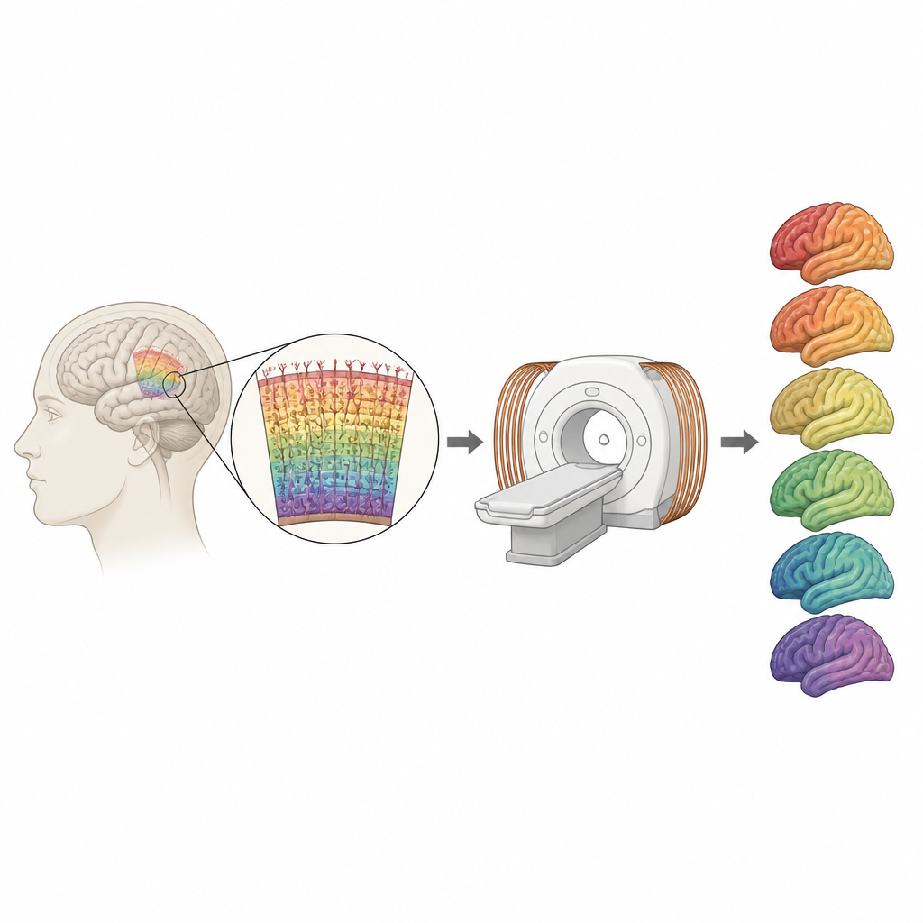
Our brains are wrapped in a thin, rippled sheet of tissue where perception, movement, and thought all arise. This outer layer, the cortex, is built from stacked layers of cells and fibers that differ from one another in subtle ways. Until now, scientists could study that fine structure only in donated brains after death. This article explains how researchers are beginning to map those layers in living people, using a new class of powerful MRI scanners and advanced analysis methods.
Why Brain Layers Matter
The cortex is not uniform. It is arranged in six main layers that differ in the size and density of nerve cells and the amount of insulated wiring, or myelin, running through them. Different regions, such as visual and motor areas, show distinct layer patterns that help shape what each region can do. For over a century, these features have been revealed by slicing and staining brain tissue under a microscope. While these classical methods gave exquisite detail, they cannot track living brains as they develop, age, or respond to disease. A key goal in modern neuroscience is to capture the same kind of layer information noninvasively, so that structure can be linked to function and clinical symptoms in real time.
A New Kind of MRI Scanner
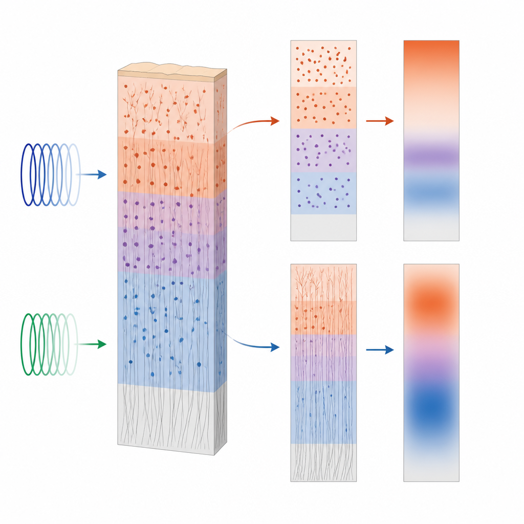
The study centers on a next-generation research MRI system called Connectome 2.0, which can generate much stronger magnetic field gradients than standard hospital scanners. These powerful gradients make diffusion MRI more sensitive to how water molecules move through tissue on microscopic scales. By applying a model known as soma and neurite density imaging, or SANDI, the team separates the signal coming from cell bodies (somas), the thin projections of nerve cells (neurites), and the surrounding space. To sharpen the view, they use a super-resolution technique that blends information from standard diffusion scans and high-quality anatomical scans, effectively pushing the diffusion data down to one millimeter resolution across the cortex.
Reading Cell Bodies and Wiring Across Depth
With these tools, the researchers sample SANDI measurements at 21 depth levels from the brain surface down to the white matter. They find that the signal linked to cell bodies peaks roughly halfway through the cortex, while the signal linked to neurites steadily climbs toward deeper layers near the white matter. These trends closely resemble patterns in histological atlases based on real tissue, where mid-depth layers are packed with large neurons and deeper layers contain dense bundles of myelinated fibers. The team also shows that sensory areas such as visual cortex differ from motor areas in how these signals vary with depth, echoing long-known differences in their cellular makeup. Even within the motor cortex, subtle changes between neighboring subregions become visible only when layer-specific measurements are examined.
Shape of the Brain and Its Microstructure
The cortex is folded into ridges and grooves, and the study reveals that the relationship between tissue structure and surface shape changes with depth. Near the surface, regions buried in grooves tend to show higher cell-body-related signal than exposed ridges. Deeper down, this pattern reverses, with ridges showing higher values than grooves. This depth-dependent flip matches earlier microscopic work on how cell density varies across folds. Together with the depth profiles of neurite signal, the results point to a rich interplay among cortical geometry, cell packing, and wiring that can now be probed in living humans.
Comparing Old and New Technology
To see what the new hardware adds, the authors compare SANDI measurements from the Connectome 2.0 scanner to those from its predecessor, Connectome 1.0, which already surpassed clinical systems. The newer scanner boosts the neurite-related signal across the cortex without altering the overall cell-body-related signal, improving sensitivity to the brain’s wiring while keeping estimates of cell bodies stable. It also reduces variability between people and better captures differences among small regions, suggesting that stronger gradients and shorter scan timings sharpen the view of both soma and neurite compartments.
What This Means for Brain Health
For non-specialists, the key message is that scientists are learning to see the fine-grained architecture of the brain’s surface in living people, at a level once reserved for microscope slides. By matching MRI-based layer profiles to trusted tissue atlases, this work shows that advanced diffusion MRI can serve as a stand-in for histology. In the future, similar methods, adapted to more widely available scanners, may help doctors and researchers track how diseases such as multiple sclerosis, dementia, or psychiatric conditions subtly alter specific layers and regions of the cortex over time.
Citation: Lee, H., Ma, Y., Chan, KS. et al. Visualizing cortical laminar architecture in the living human brain using next-generation ultra-high-gradient diffusion MRI. Commun Biol 9, 651 (2026). https://doi.org/10.1038/s42003-026-09887-2
Keywords: cortical layers, diffusion MRI, brain microstructure, Connectome 2.0, SANDI model