Clear Sky Science · en
Cortical gradient compression links cognition and transcriptomic signatures in primary angle-closure glaucoma
When Eye Disease Reaches the Brain
Primary angle closure glaucoma is best known as an eye disease that raises pressure inside the eye and can lead to blindness. This study shows that the condition also reshapes how the brain is organized and may help explain why some patients experience not only vision loss but also problems with memory and thinking.
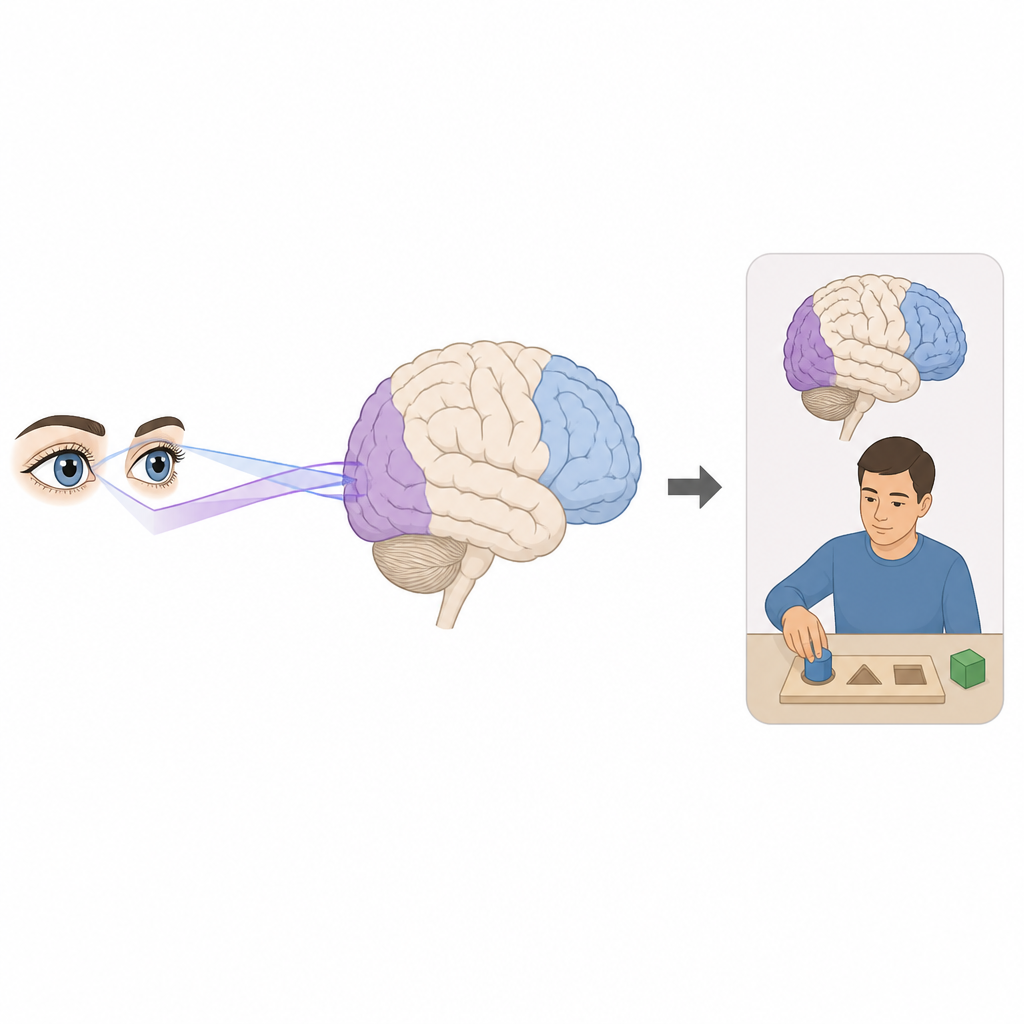
From Blurry Vision to Thinking Problems
Primary angle closure glaucoma blocks the normal drainage of fluid from the front of the eye, damaging the optic nerve and narrowing the visual field. Doctors have long suspected that this damage does not stop at the eye. People with the disease can show signs of cognitive decline similar to those seen in dementia. Yet it has been unclear how changes in brain organization might connect vision loss to difficulties with attention, memory, and other mental abilities.
Mapping Brain Highways at Rest
To probe this link, researchers scanned the brains of 66 people with primary angle closure glaucoma and 69 similar adults without the disease while they rested quietly in an MRI scanner. Instead of looking at single brain regions, they used a method called gradient mapping that captures how activity patterns blend smoothly from areas that handle simple sensations, like sight and movement, toward regions that support daydreaming, self reflection, and complex thought. These gradual shifts can be thought of as the brain’s main highways, organizing how information flows from seeing to understanding.
Compressed Brain Gradients and Network Shifts
The scans revealed that these brain highways were compressed in people with glaucoma. The spread of gradient values that normally separates basic visual areas from higher thinking regions was narrowed, suggesting that different parts of the cortex had become less distinct in their roles. This effect appeared across many networks, but it was especially strong in the visual system and in the default mode network, which is active when the mind is at rest and engaged in internal thoughts. In the visual network, stronger compression tracked with worse visual acuity, and in the default mode network, a more extreme shift in position was linked to lower scores on a standard test of overall cognition. Together, these results suggest that disrupted brain hierarchy may sit between eye damage and the thinking problems some patients experience.
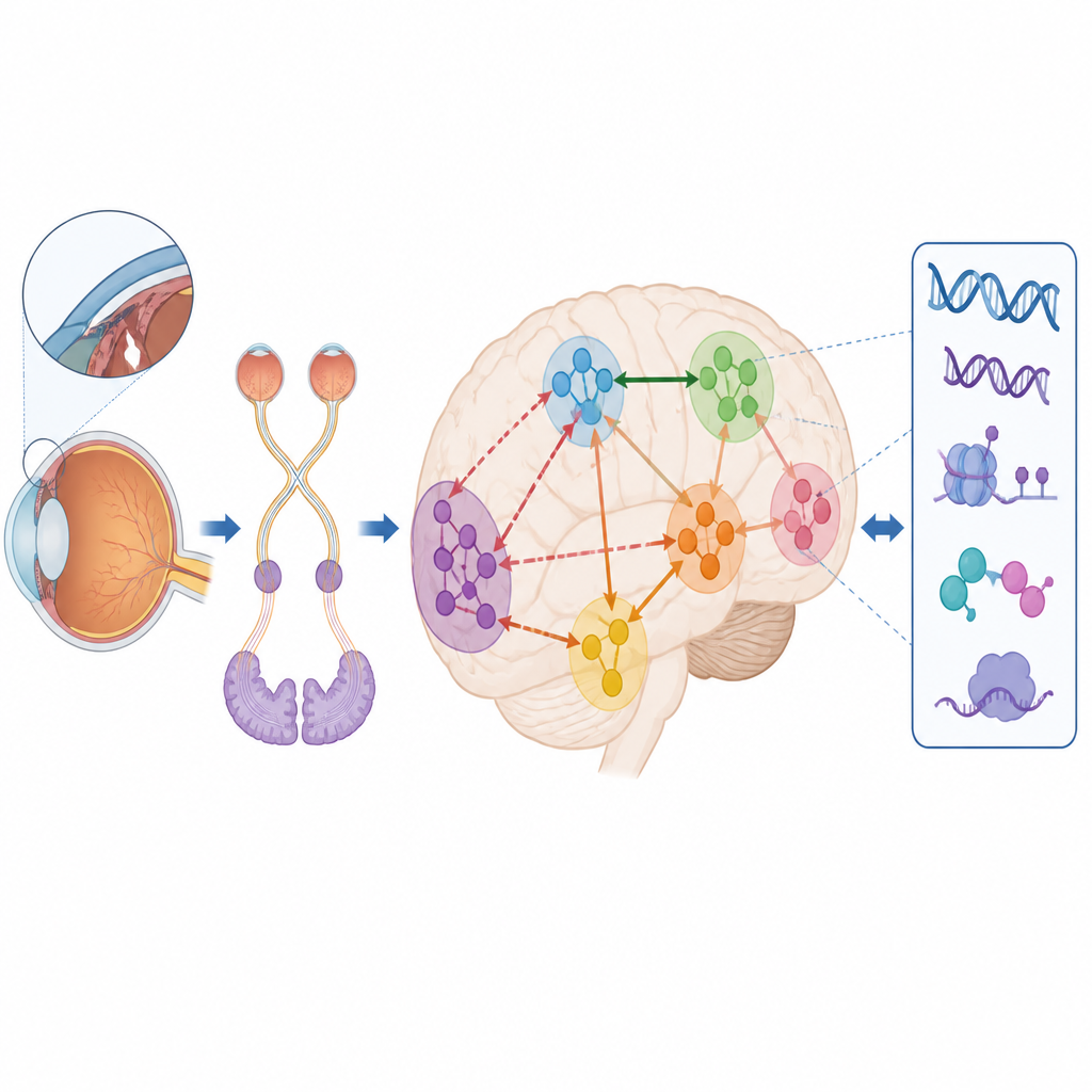
Clues from the Brain’s Genetic Blueprint
The team then asked whether these large scale changes in brain organization might reflect deeper molecular patterns. They used a public atlas of gene activity measured in brain tissue from donors without glaucoma and compared it with the pattern of gradient changes seen in the patients. Thousands of genes showed expression patterns that lined up with the altered gradients, especially in the back of the brain where visual processing begins. Genes most closely tied to the disruptions were involved in how nerve cells signal to each other, how developing brain circuits are shaped, and how support cells called astrocytes help maintain healthy networks. This points to a possible chain that runs from genes and cell types, through brain networks, to behavior.
What This Means for Patients and Future Work
In simple terms, the study suggests that primary angle closure glaucoma is not only a disease of the eye but also a disorder of how the brain’s communication scaffolding is arranged. As vision declines, the normal separation and balance between seeing regions and thinking regions becomes squeezed, and this tighter, less flexible layout is tied to worse visual and cognitive performance. While the work is cross sectional and cannot yet say which changes come first, it offers a new way to understand why some patients struggle with memory and attention and hints at molecular pathways that future therapies might target.
Citation: Wang, Y., Liang, X., Yang, C. et al. Cortical gradient compression links cognition and transcriptomic signatures in primary angle-closure glaucoma. Commun Biol 9, 653 (2026). https://doi.org/10.1038/s42003-026-09886-3
Keywords: primary angle closure glaucoma, brain networks, cognitive impairment, resting state fMRI, gene expression