Clear Sky Science · en
Perinatal liver sympathetic innervation governs body size
How Early Nerves Help Set Our Final Size
Why do some children grow more slowly, even when their hormones look normal? This study explores a surprising answer: in the days around birth, tiny nerve fibers that connect the brain to the liver quietly help decide how big we become. When this nerve wiring goes wrong, the liver makes less of a key growth signal, stunting the body even if classic growth hormones are present. Understanding this hidden nerve–liver link could open new ways to think about growth problems and some neurodevelopmental conditions.

The Usual Path from Brain to Body Growth
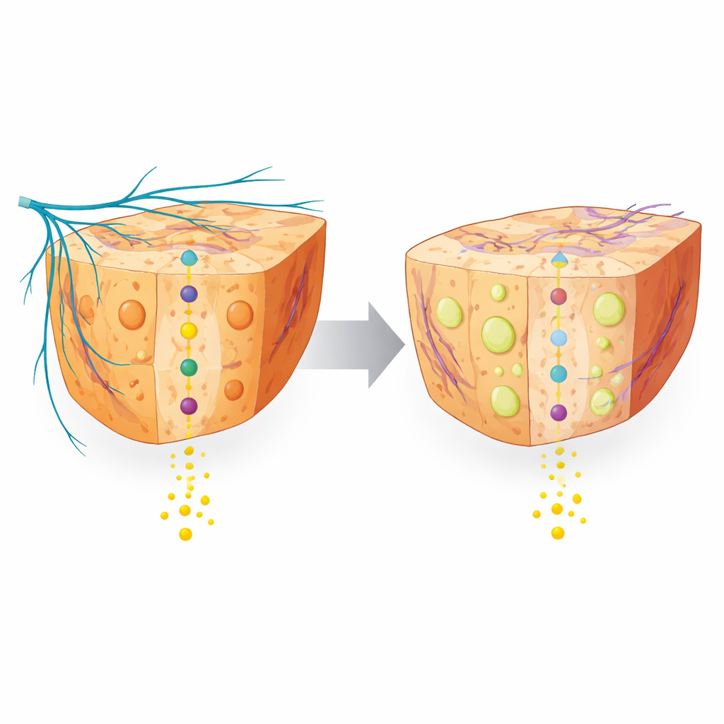
Body growth in childhood is usually explained by a simple chain: the brain releases a messenger that tells the pituitary gland to send out growth hormone, which then travels through the blood to the liver. In response, the liver produces insulin-like growth factor 1, or IGF‑1, a protein that promotes cell growth in many tissues and strongly influences final body size. Most medical approaches to poor growth focus on fixing this hormone chain. But the liver is also wired to the nervous system, especially to sympathetic nerves that carry signals from the brain and spinal cord. The authors wondered whether problems in early brain development might disturb this wiring and, through the liver, slow growth in a way that standard hormone tests could miss.
Clues from a Child and from Nerve-Damaged Newborn Mice
The story began with a child carrying a harmful mutation in a gene called Cdh1, who showed microcephaly (a small brain), delayed development, and poor weight gain. When the team measured his blood, they found that his IGF‑1 and a major IGF‑1 binding partner were well below the normal range for his age, hinting that the liver’s growth signal was weakened. To probe this connection in a controlled way, the researchers first used newborn mice and chemically injured their sympathetic nerves just after birth. These pups quickly developed reduced nerve fibers in the liver, grew more slowly, and showed greatly reduced activity of genes required to make IGF‑1 and its stabilizing partners. Notably, this change was strongest in the liver, suggesting that this organ is especially dependent on intact sympathetic wiring in the perinatal period.
A Mouse Model Linking Brain Development to Liver Wiring
Next, the team created mice lacking Cdh1 only in nerve cells, starting late in embryonic life. At birth, these animals looked normal, but between one and three weeks after birth they fell behind their littermates in body weight. Their hearts, lungs, and kidneys were relatively spared, but their livers were small, and detailed imaging revealed immature brain structures and unstable neural connections. In the liver, overall nerve density and particularly sympathetic fibers were markedly reduced. The animals also lost muscle and fat tissue and showed weaker grip strength, signs consistent with widespread effects of sympathetic malfunction. Yet, the brain–pituitary side of the classic growth pathway appeared intact: the numbers and shapes of key hypothalamic neurons, blood levels of growth hormone–releasing hormone and growth hormone, and growth hormone–producing pituitary cells were all normal.
How Miswired Nerves Disturb the Liver’s Growth Signal
With the usual hormones in place, the researchers looked inside liver cells to see where the chain broke. Growth hormone still bound its receptor and triggered an early enzyme step normally, but a crucial downstream protein, STAT5, was less activated. STAT5 is needed to switch on the IGF‑1 gene, so its failure explained the low IGF‑1 output. Both the nerve-damaged newborn mice and the Cdh1-deficient mice showed similar defects at this point in the pathway. At the same time, their livers accumulated excess fat droplets and glycogen and began to develop scarring, changes known to disturb the behavior of surface receptors and signaling molecules. Indeed, the physical interaction between STAT5 and its upstream partner was weakened. In effect, faulty sympathetic wiring pushed the liver into an unhealthy metabolic state that blocked the final step needed to make IGF‑1.
Rescuing Growth and What It Means for Children
To test whether boosting the missing growth signal could overcome the wiring defect, the team gave young Cdh1-deficient mice injections of IGF‑1 during the second week of life. This simple treatment restored their body weight, normalized the ratio of brain to body size, and partly corrected liver size, even though it did not repair the damaged liver nerves or fix IGF‑1 production inside the organ. The work shows that an early neurodevelopmental problem can indirectly stunt growth by disrupting liver nerve connections, independently of the usual growth hormone pathway. For lay readers, the key message is that healthy growth is not governed by hormones alone: it also depends on proper nerve wiring between the brain and organs like the liver. In some children with neurodevelopmental disorders, looking beyond classic hormone tests to consider this nerve–liver axis—and carefully timed IGF‑1 support—may one day offer new options for improving growth.
Citation: Bobo-Jimenez, V., Gomila, S., Lapresa, R. et al. Perinatal liver sympathetic innervation governs body size. Commun Biol 9, 596 (2026). https://doi.org/10.1038/s42003-026-09880-9
Keywords: growth hormone, liver innervation, IGF-1, neurodevelopment, body growth