Clear Sky Science · en
Periplasmic crowding and peptidoglycan hydrolase activity as drivers of outer membrane vesiculation in Acinetobacter baumannii
How Bacteria Throw Out Their Trash
Antibiotic resistant bacteria have many tricks for staying alive in hostile environments, including ways to dump damaged parts of themselves before those parts cause harm. This study looks at one such strategy in the hospital pathogen Acinetobacter baumannii, showing how it forms tiny bubbles from its outer surface to shed waste and relieve internal stress, a process that may influence how infections spread and resist treatment.
Tiny Bubbles on a Bacterial Skin
Many harmful bacteria are wrapped in a double outer skin. From this surface they release minuscule bubbles, called outer membrane vesicles, that pinch off and float away. These bubbles can carry bits of the outer surface, toxins, and even genetic material, helping bacteria share resistance genes, talk to neighbors, and interact with host tissues. Yet scientists still lack a clear picture of what actually pushes the cell surface to bulge outward and snap off these bubbles.
When the Space Between Layers Gets Crowded
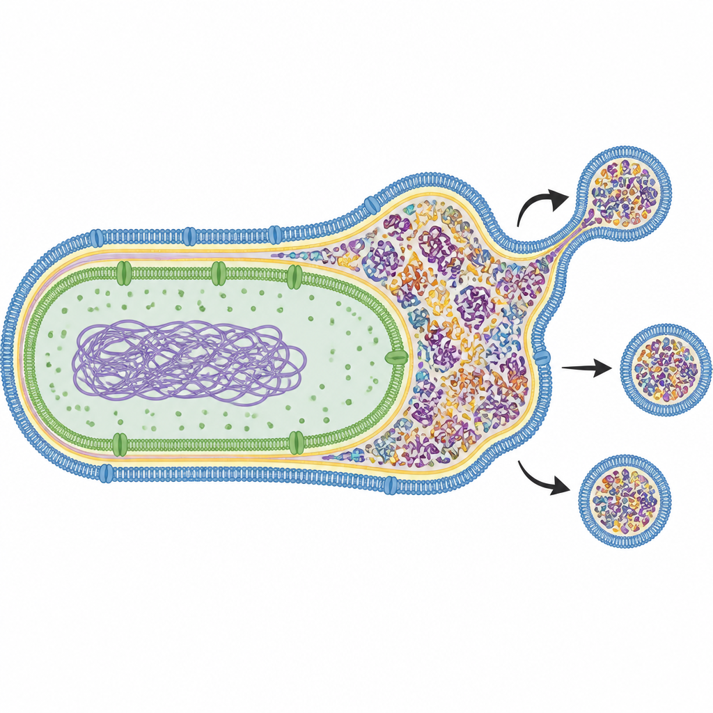
The team focused on the slim compartment between the inner and outer skins of the cell, a region packed with important proteins. They studied a mutant strain missing DegP, a protein that normally acts as both a helper and a cleanup enzyme for misfolded proteins, especially at high temperature. When this safety valve is removed and the cells are warmed, misfolded proteins and bits of cell wall material build up in the narrow space. Using a technique that tracks the movement of glowing proteins, the researchers showed that this space becomes so crowded that molecules can no longer move freely, a sign of strong internal pressure. At the same time, the cells began releasing many more outer membrane vesicles than normal.
Leaky Skin Is Not Enough
The scientists then asked whether simply damaging the outer skin would cause more bubble release. They compared different mutants that each disturb surface proteins in their own way and measured how easily dyes, antibiotics, and a detergent like bile salt could enter. Some strains, such as those lacking the chaperone SurA, had very leaky outer membranes but still produced few vesicles. In contrast, the DegP mutant showed both increased leakiness and a dramatic rise in vesicle production. Careful imaging with electron microscopes revealed that, in this mutant, the distance between the inner and outer skins widened and the surface sprouted tubular protrusions. These findings suggested that weakened skin alone cannot explain vesicle formation; something else inside the wall must also change.
Cutting the Cell Wall to Release the Pressure
Attention turned to enzymes that trim and recycle the sugar mesh making up the rigid cell wall. Protein surveys of the vesicles from DegP mutants uncovered high levels of lytic transglycosylases, especially MltB and MltD, and an amidase called AmiAb, all of which cut cell wall building blocks into smaller fragments. Chemical analysis of the cell wall showed that the DegP mutant accumulated unusual fragments, pointing to extra cutting activity. Under the microscope, these cells produced more and larger vesicles, some with a double shell that wrapped both the inner and outer membranes, and the vesicle interiors were enriched in sugars derived from the cell wall. When the researchers deleted the mltB gene, vesicle formation in the DegP mutant was nearly abolished and the cells died sooner under heat stress, implying that controlled cutting of the cell wall is needed to bud off vesicles and survive.
Two Conditions Needed for Bubble Making
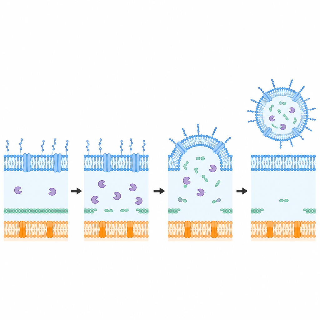
To test whether crowding and wall cutting must work together, the team overproduced the MltB enzyme in a strain that had a leaky outer skin but normally made few vesicles. In this setting, boosting MltB triggered clear bulges and vesicle-like structures on the surface. Across many experiments, a consistent picture emerged: only when the space between the membranes is jammed with misplaced proteins and fragments, and when cell wall cutting enzymes are active, does the outer surface bend and pinch off robustly into vesicles. If the wall cutting is blocked, pressure builds but bubbles do not form efficiently; if the wall is cut without strong crowding, the response is weaker.
Why This Matters for Infections
For a non-specialist, the takeaway is that Acinetobacter baumannii uses a two-step safety release system to cope with stress at high temperature and other harsh conditions. First, the failure of DegP allows the space between its inner and outer skins to fill up with misfolded proteins and cell wall scraps, creating pressure. Second, wall-cutting enzymes such as MltB and MltD loosen the rigid mesh, allowing the outer skin to bulge out and shed loaded vesicles that carry away the excess material. This coupling between inner crowding and controlled weakening of the wall helps the bacteria maintain their surface, survive stress, and potentially shape how they respond to antibiotics and the immune system.
Citation: Kim, B., Son, Y., Lee, R. et al. Periplasmic crowding and peptidoglycan hydrolase activity as drivers of outer membrane vesiculation in Acinetobacter baumannii. Commun Biol 9, 617 (2026). https://doi.org/10.1038/s42003-026-09876-5
Keywords: outer membrane vesicles, Acinetobacter baumannii, bacterial cell wall, envelope stress, antibiotic resistance