Clear Sky Science · en
Simulated closed-loop magnetic stimulation promotes function recovery and axonal regeneration in spinal cord injury
Helping the Injured Spine Talk to the Brain Again
Spinal cord injury often means a sudden loss of movement and independence, with few options for restoring function. This study explores a noninvasive way to "coach" damaged nerve pathways back to life using carefully timed magnetic pulses applied outside the body. By synchronizing stimulation of the brain and lower spinal nerves in mice, the researchers show that it is possible not only to improve walking but also to regrow key nerve fibers through injured spinal tissue.
A New Kind of Paired Magnetic Therapy
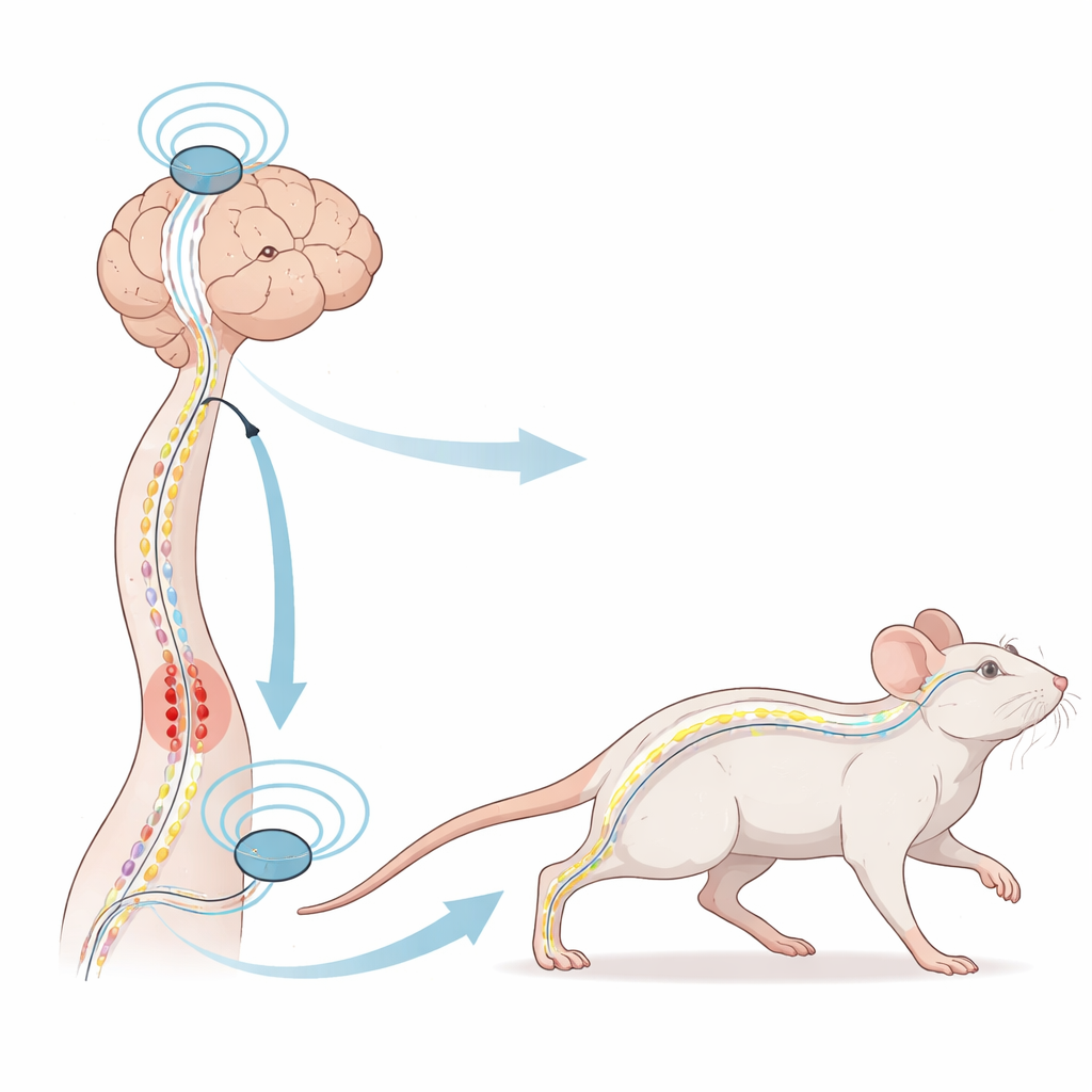
The team developed what they call simulated closed-loop magnetic stimulation, or SCMS. Instead of stimulating just the brain or just the spinal nerves, SCMS delivers pulses to both ends of the movement pathway in a tightly timed sequence. One coil sits over the motor region of the brain that sends signals down the spinal cord, while a second coil targets a nerve root near the lower back that carries sensory information upward. By matching the timing of these two inputs to the natural speed of nerve conduction, SCMS is designed to mimic the normal back-and-forth dialogue between brain and limbs that is disrupted after injury.
Testing Movement Recovery in Mice
To see whether this approach could restore function, the researchers created precise spinal cord injuries in mice and compared three groups: injured animals with no treatment, animals receiving standard brain-only magnetic stimulation, and animals receiving the new SCMS protocol. Over six weeks, they filmed the mice walking and used detailed gait analysis software to track step patterns, balance, and speed. They also recorded electrical activity from leg muscles and examined muscle tissue under the microscope. Mice treated with SCMS showed markedly better hind-limb coordination, higher foot lift, more symmetrical posture, and stronger, more frequent muscle contractions than untreated or brain-only stimulated animals. Their leg muscles were less wasted, and more of the special nerve–muscle contact sites, called neuromuscular junctions, were fully restored.
Following Signals Through the Injured Spinal Cord
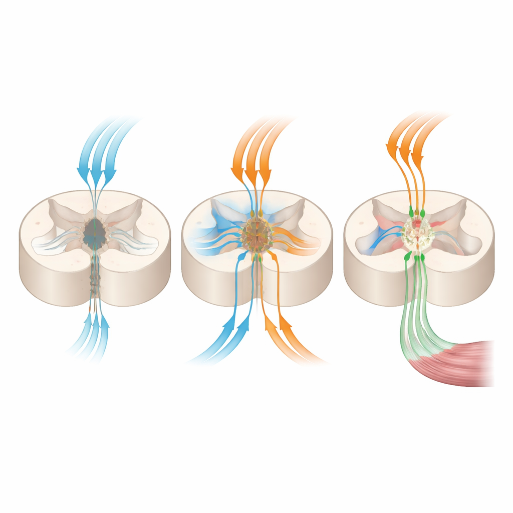
Improved walking could come from many sources, so the team directly measured how well signals traveled through the damaged spinal cord. Using implanted optical fibers and fluorescent calcium sensors that light up when neurons fire, they tracked activity in brain cells that send movement commands and in spinal cord regions where these commands arrive. In untreated or brain-only stimulated mice, signal strength in these pathways dropped sharply after injury. In contrast, SCMS-treated mice showed much stronger activation both in the motor cortex and in corticospinal axons below the injury site. Electrical tests confirmed that motor-evoked responses traveled more reliably from brain to leg muscles only when SCMS was applied, suggesting that the main movement highway—the corticospinal tract—had been partially rebuilt.
Regrowing Nerve Fibers and Rebuilding Circuits
To check for actual regrowth of nerve fibers, the scientists labeled corticospinal tract axons with a fluorescent marker and examined the injured spinal cords. In most injured mice, these fibers stopped abruptly at the scar border and did not cross into tissue below the lesion. In SCMS-treated animals, however, some axons sprouted through the scar and extended up to about two millimeters beyond the injury, a notable feat in adult central nervous system tissue where regeneration is usually extremely limited. These regenerated fibers formed contacts with specific spinal "middleman" neurons involved in controlling movement and connected onward toward muscles, indicating that SCMS helped rebuild functional sensorimotor circuits rather than just random growth.
Turning On the Nerve’s Own Repair Programs
Finally, the researchers asked what internal repair programs were being activated in the spinal cord. They combined large-scale measurements of gene activity, proteins, and small molecules in tissue from treated and untreated mice. Across all three data types, one pathway stood out: a metabolic and growth-control route centered on the enzyme AMPK and its partners CREB and BDNF. SCMS boosted this pathway and increased levels of BDNF, a well-known nerve growth factor that supports neuron survival and axon extension. Blocking or failing to engage such routes in other studies has limited recovery, so their activation here offers a plausible explanation for how timed magnetic stimulation can encourage damaged axons to regrow and form useful connections.
What This Could Mean for People
In plain terms, this work shows that a carefully choreographed pattern of magnetic pulses applied to the head and lower back can help an injured spinal cord in mice reconnect with the brain, restore more natural walking, and even regrow crucial nerve fibers through scarred tissue. The treatment is noninvasive and uses stimulation strengths similar to those already considered safe in human clinics. While much research remains before it can be applied to patients, especially to confirm long-term safety and precise wiring of new connections, SCMS points toward a future in which external devices may guide the body’s own repair machinery to rebuild broken communication lines after spinal cord injury.
Citation: Zhang, L., Xiao, Z., Xia, C. et al. Simulated closed-loop magnetic stimulation promotes function recovery and axonal regeneration in spinal cord injury. Commun Biol 9, 614 (2026). https://doi.org/10.1038/s42003-026-09848-9
Keywords: spinal cord injury, magnetic stimulation, axon regeneration, corticospinal tract, neurorehabilitation