Clear Sky Science · en
Stem cells resume asymmetric division upon niche re-entry through reactivating the centrosome orientation checkpoint
Why stem cells don’t lose their sense of direction
Our tissues rely on stem cells to keep working for a lifetime, yet stem cells themselves can be lost or damaged. This study in fruit flies explores how replacement stem cells regain the ability to divide in a precise, one‑in, one‑out manner that prevents tissue overgrowth or depletion. By watching living testes over many hours, the authors reveal how cells that step back from a more mature state re‑enter the stem cell “niche” and rapidly recover a built‑in quality‑control system that keeps their divisions properly oriented.
A tiny factory at the tip of the testis
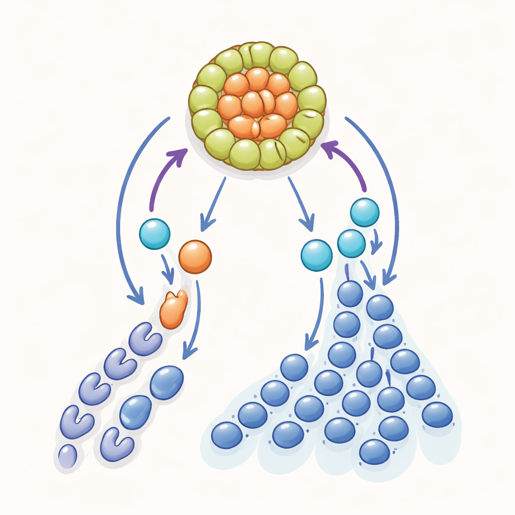
In the male fruit fly, sperm are produced in a long, coiled tube. At its tip sits a small cluster of support cells known as the hub, surrounded by 8–12 germline stem cells. Each stem cell normally divides asymmetrically: the side touching the hub stays a stem cell, while the opposite side becomes a daughter cell that moves away and begins the journey toward becoming sperm. This simple geometric rule—one daughter stays, one leaves—keeps the stem cell pool stable while constantly supplying new sperm precursors.
When mature cells turn back the clock
Despite this careful balance, stem cells do occasionally leave the hub or are lost with age and stress, so the system needs a backup plan. Earlier work showed that lost stem cells can be replaced in two ways: by “symmetric renewal,” where both daughters of a division stay in the niche, or by “dedifferentiation,” where a more advanced germ cell moves back toward the hub and regains stem cell traits. Using gentle, long‑term live imaging that spanned roughly 1,400 hours of observation, the authors tracked hundreds of divisions in normal testes and in testes recovering from an experimental depletion of stem cells. They found that most replenishment came from dedifferentiation events, particularly from single gonialblasts—the immediate daughters of stem cells—migrating back and reattaching to the hub.
A cell‑cycle pause before rejoining the niche
Surprisingly, these returning cells did not behave like “blank slates.” Once they reattached to the hub, they almost always divided very soon afterward—on average just over two hours later, compared with a full stem cell cycle of about 14 hours. Yet they did not keep cycling unusually fast; a second division was not seen within the next nine hours of monitoring. This timing pattern suggested that dedifferentiating cells were already parked at a specific stage of the cell cycle before they reached the niche. By using a fluorescent cell‑cycle reporter, the team showed that, at the moment of reentry, all observed returning cells were in late G2, the checkpoint just before a cell commits to mitosis. In other words, these cells seem to wait in a poised state and then complete division shortly after regaining contact with the hub.
Reawakening a built‑in direction sensor
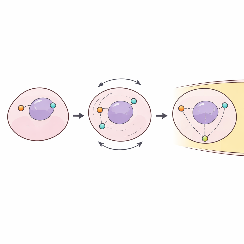
The authors next asked why these poised cells sit in late G2. In normal stem cells, a specialized quality‑control system called the centrosome orientation checkpoint monitors whether the internal division machinery is lined up perpendicularly to the hub. If not, the cell is held in late G2 until its two organizing centers—the centrosomes—are correctly positioned, ensuring that one daughter will remain in the niche and the other will leave. Tests with a microtubule‑disrupting drug confirmed that this checkpoint is usually active only in stem cells, not in their more mature daughters. However, during niche regeneration, a significant fraction of daughter cells outside the hub started to show signs of this checkpoint, implying that it had been reactivated as they prepared to dedifferentiate. When a key polarity protein, Bazooka/Par‑3, required for this checkpoint, was knocked down, the recovery of stem cell numbers after depletion was noticeably slowed, suggesting that the checkpoint helps make dedifferentiation efficient and safe.
Snapping the internal compass back into place
Live imaging of centrosomes provided a final piece of the puzzle. As soon as a dedifferentiating cell reattached to the hub, one centrosome rapidly moved—or was already positioned—toward the hub‑facing side of the cell, while the other shifted to the opposite side, typically within about half an hour. This swift reorientation, followed by timely mitosis, means that replacement stem cells quickly regain the same directional division pattern as native ones. The study also shows that dedifferentiated stem cells derived from very early daughters (gonialblasts) have much better orientation control than those derived from later, multi‑cell stages, underscoring that the source stage matters.
How tissues keep balance over a lifetime
Together, these findings reveal that when more mature germ cells in the fly testis are called back into stem cell duty, they do not simply move into the right place; they also switch on a stem cell–specific checkpoint that pauses them until their internal compass is reset. This coupling of dedifferentiation to a polarity checkpoint allows the niche to refill itself without sacrificing the precise one‑in, one‑out division pattern that prevents overgrowth or depletion. Because similar principles of niches, checkpoints, and dedifferentiation are found in other tissues and organisms, this work offers a window into how our own stem cells may preserve order while adapting to damage and aging.
Citation: Bener, M.B., Twillie, A., Patel, N. et al. Stem cells resume asymmetric division upon niche re-entry through reactivating the centrosome orientation checkpoint. Commun Biol 9, 556 (2026). https://doi.org/10.1038/s42003-026-09812-7
Keywords: stem cell niche, dedifferentiation, asymmetric division, Drosophila testis, cell cycle checkpoint