Clear Sky Science · en
Structural basis for antibody cross-neutralization of Dengue and Zika viruses
Why this matters for future vaccines
Mosquito-borne diseases like dengue and Zika can turn a single bite into a life‑threatening illness, and attempts to make vaccines have stumbled because the wrong kind of antibodies can actually make a second infection worse. This study takes a close, atomic‑level look at a rare “good” antibody that safely shuts down both dengue and Zika viruses. By understanding exactly how this antibody latches onto the viruses, scientists hope to design smarter vaccines that protect against many related viruses at once without backfiring.
A closer look at dangerous cousins
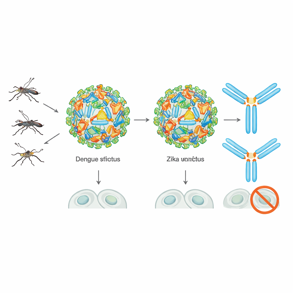
Dengue virus comes in four flavors, or serotypes, and together they are estimated to infect hundreds of millions of people every year. Zika, once obscure, exploded across the Americas a decade ago, linked to birth defects and neurological disease. These viruses belong to the same family and are wrapped in a coat of envelope proteins that mediate entry into human cells and are the main targets for antibodies. The immune system’s response is complicated: antibodies from a past dengue or Zika infection can sometimes help a different dengue serotype infect cells more efficiently, a phenomenon called antibody‑dependent enhancement. That history has made vaccine developers deeply wary, especially after safety concerns derailed the first licensed dengue vaccine.
Discovering a broadly protective antibody
Among the many antibodies people make after infection, a few rare ones can neutralize all four dengue serotypes and often Zika as well. Earlier work had identified a group called EDE1 antibodies, which recognize a site formed where two envelope proteins meet as a dimer on the virus surface. Recently, the authors’ team found another broadly neutralizing antibody, named F25.S02, that potently blocks dengue 1–4 and Zika but targets this region in a subtly different way. In this study, they set out to visualize, in three dimensions, exactly how F25.S02 engages its target on both viruses to understand what makes it so broadly effective.
Seeing the binding site in atomic detail
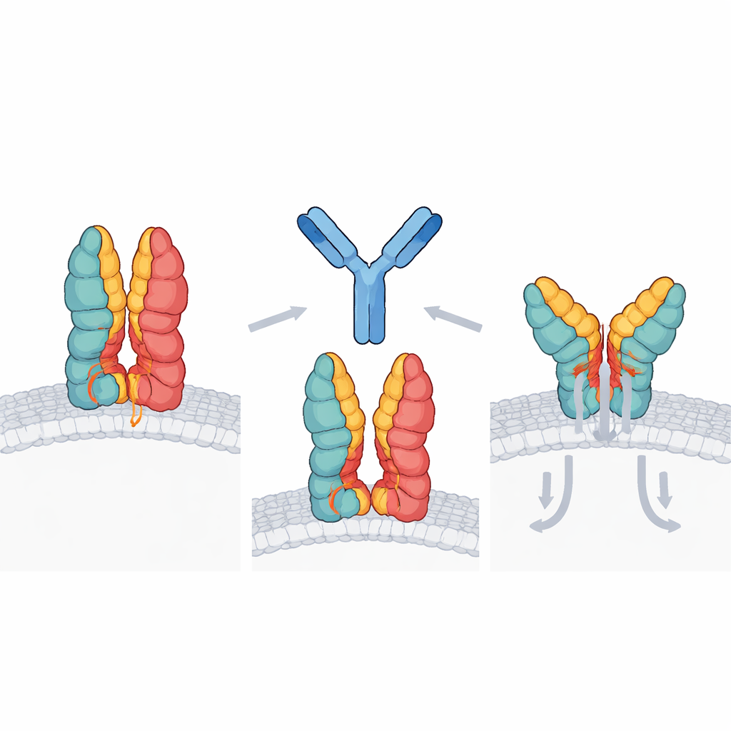
The researchers used cryo‑electron microscopy to image F25.S02 bound to a stabilized version of the dengue type 3 envelope protein dimer, and X‑ray crystallography to capture the antibody paired with the Zika envelope dimer. Both approaches revealed the same overall picture: two copies of F25.S02 clamp onto each envelope dimer at a highly conserved patch spanning the interface of the two protein partners. The antibody interacts most strongly with a region called the fusion loop in one partner along with portions of adjacent domains in both partners—exactly the parts that must move and reorganize when the virus fuses with a host cell membrane. Strikingly, almost all of the gripping force comes from the antibody’s heavy chain, while the light chain plays only a minor supporting role.
A distinct way of locking the virus shut
Comparing F25.S02 with previously known EDE1 antibodies revealed important differences that could matter for vaccine design. EDE1 antibodies tend to sit closer to the two‑fold symmetry axis of the envelope dimer and share the binding work fairly evenly between their heavy and light chains. F25.S02, in contrast, is shifted slightly away from the center and relies overwhelmingly on its heavy chain, with most of its contact points encoded directly by its original, or germline, gene sequence. The antibody can tolerate sugar decorations (glycans) on the viral surface that lie within its footprint, adjusting around them without depending on them for binding. Modeling its position on an intact Zika particle suggests that many copies could bind without bumping into each other, cross‑linking dimers and restricting the shape changes needed for fusion.
What this means for next‑generation vaccines
For non‑specialists, the key takeaway is that this work maps a shared “Achilles’ heel” on both dengue and Zika viruses and shows how one natural antibody can exploit it with minimal fine‑tuning. Because the targeted patch is highly conserved across all four dengue types and Zika, and because the antibody’s heavy chain already fits that site well in its near‑germline form, it should be feasible to design vaccine proteins that specifically present this vulnerable interface and encourage the immune system to make F25.S02‑like antibodies. Such structure‑guided vaccines could offer broad protection while avoiding the risky, infection‑enhancing antibodies that have hampered past efforts, bringing us closer to safe, durable defenses against multiple mosquito‑borne viruses.
Citation: Hurlburt, N.K., Lubow, J., Goo, L. et al. Structural basis for antibody cross-neutralization of Dengue and Zika viruses. Commun Biol 9, 568 (2026). https://doi.org/10.1038/s42003-026-09805-6
Keywords: dengue, Zika, broadly neutralizing antibodies, vaccine design, envelope protein