Clear Sky Science · en
Integrative structural insights into the IgG-FcRn interactions revealed by engineered FcRn-immobilized affinity chromatography
Why antibody lifetimes in the body matter
Many of today’s most important medicines are monoclonal antibodies used to treat cancer, autoimmune diseases, and other chronic conditions. How long these antibodies stay in a patient’s bloodstream strongly influences how well they work, how often they must be injected, and how much they cost. This paper explores a key cellular recycling system that protects antibodies from breakdown, and introduces a new experimental tool that helps scientists tune and assess that protection more precisely.
A cellular recycling gatekeeper
Our bodies rely on a protein called the neonatal Fc receptor, or FcRn, to rescue antibodies from being destroyed inside cells. Antibodies are constantly taken up into tiny internal sacs, where the environment is acidic. At this low pH, FcRn grabs onto antibodies and diverts them away from cellular “garbage disposals,” returning them to the bloodstream when they reach a neutral pH again. The strength and pH dependence of this handshake between antibody and FcRn is one of the main reasons antibodies can circulate for weeks. Yet measuring this interaction cleanly and understanding exactly which parts of an antibody influence it have been surprisingly difficult.
Building a tougher test column
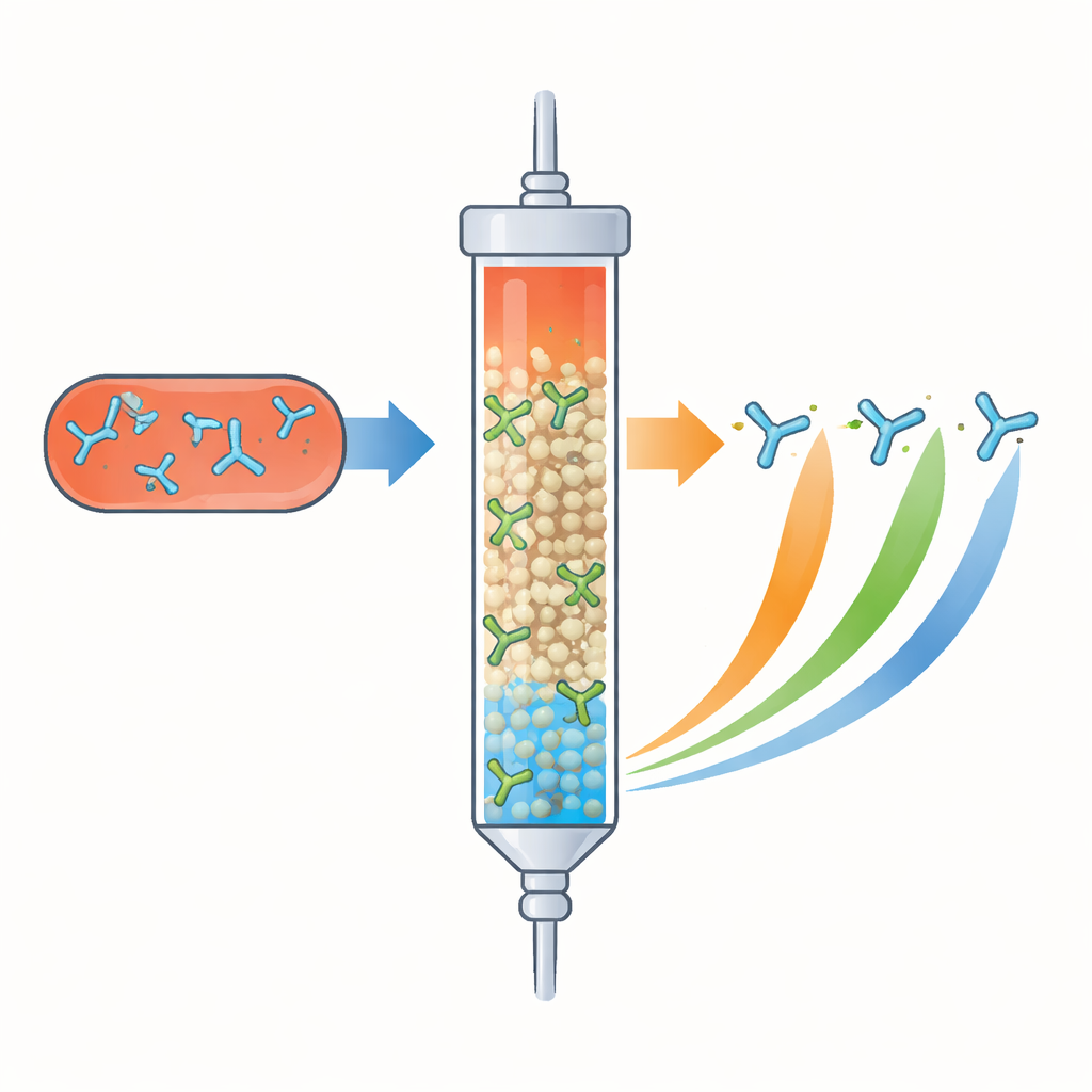
The authors tackled this challenge by engineering a robust laboratory column coated with FcRn. In affinity chromatography, such a column acts like a filter that briefly holds onto molecules that bind its surface. Here, antibodies are passed through a column where FcRn is immobilized on resin beads, and the acidity of the flowing buffer is gradually changed. Antibodies that bind FcRn more strongly or over a broader pH range cling longer and come out later, while weak binders flow through quickly. To withstand high pressures and repeated exposure to different pH and salt conditions, the team subtly altered FcRn’s own sequence to increase its thermal stability without disturbing its antibody-binding site.
Reading antibody quality from how they flow
With this engineered FcRn column, the scientists examined several types of antibodies. They first oxidized specific methionine residues known to damage FcRn binding. As the level of oxidation increased, the chromatograms split into earlier, broader peaks, clearly separating more and less damaged molecules in a single run. Next, they tested a panel of antibodies with well-studied mutations in the constant “Fc” region that are designed to raise or lower FcRn affinity. Variants with enhanced binding emerged from the column later, while a mutant that barely interacts with FcRn passed through almost immediately. These results showed that the column method not only detects FcRn affinity but can also reveal mixtures of molecules with different behaviors that are hard to parse with traditional surface-based techniques.
Surprising influence of the light chain
Beyond known Fc changes, drug developers have observed that swapping the variable “arms” of antibodies can unexpectedly alter FcRn binding and pharmacokinetics. To dissect this, the authors analyzed 13 approved IgG1 therapeutic antibodies and compared their flow through the FcRn column with the predicted electric charge (isoelectric point) of different regions. They found only modest links to the heavy chain and overall molecule, but a stronger association with the light chain and its variable region, especially in areas outside the classical antigen-binding loops. Focusing on adalimumab as a model antibody, they introduced specific positive or negative charges at three positions on the side surface of the light chain. Small negative changes made the antibody elute earlier (weaker FcRn binding), while adding a positive charge delayed elution (stronger binding). Independent measurements with surface plasmon resonance confirmed these shifts in affinity.
A structural picture of charge and recycling
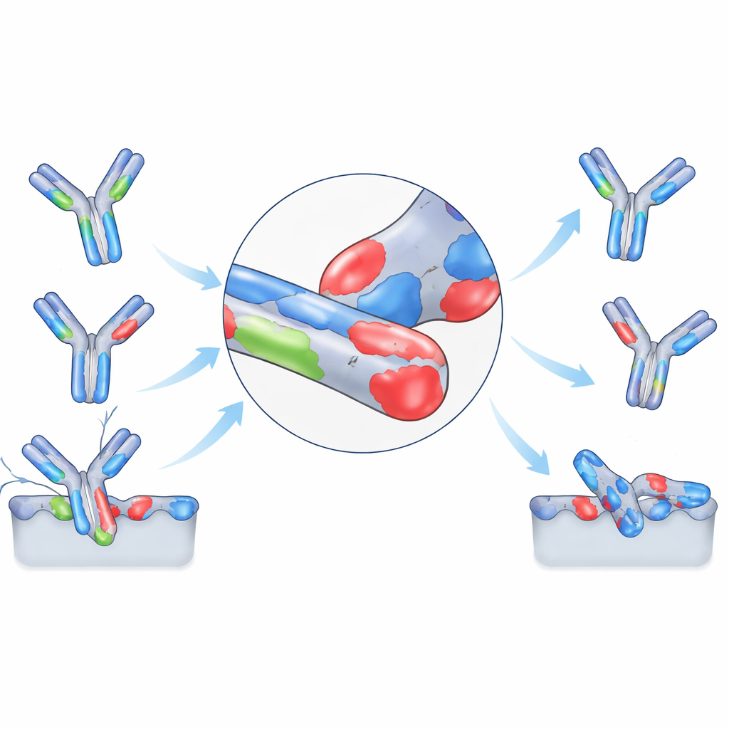
To interpret these findings, the researchers assembled a three-dimensional model combining known crystal structures of a full antibody, FcRn, and human serum albumin on a membrane surface. In their favored “reclined” arrangement, two FcRn molecules engage the antibody’s Fc region, while the antibody’s arms lie near the cell membrane. In this configuration, the lateral face of the light chain comes close to a patch of negatively charged residues on FcRn and its partner protein, β2-microglobulin. Positive charges on the light chain can therefore strengthen the interaction, whereas added negative charges weaken it. At the same time, if an antibody becomes too positively charged overall, it may stick nonspecifically to cell surfaces and be cleared faster, so there is a delicate balance to strike.
What this means for future antibody medicines
To a non-specialist, the takeaway is that the authors have created a more realistic and discriminating way to “test-drive” antibody recycling in the lab, and used it to uncover how subtle charge patterns on the antibody’s light chain help tune that process. Their FcRn column can separate good from poor binders, spot chemical damage, and reveal how particular design tweaks alter recycling-friendly interactions. Although translating these measurements into exact lifetimes in patients remains complex, the structural insights and practical assay described here should help drug developers design antibody therapies that last longer, work more reliably, and are easier to characterize for quality and safety.
Citation: Kiyoshi, M., Suzuki, T., Inoue, N. et al. Integrative structural insights into the IgG-FcRn interactions revealed by engineered FcRn-immobilized affinity chromatography. Commun Biol 9, 513 (2026). https://doi.org/10.1038/s42003-026-09789-3
Keywords: therapeutic antibodies, neonatal Fc receptor, antibody pharmacokinetics, protein engineering, affinity chromatography