Clear Sky Science · en
Downfield magnetic resonance signals serve as endogenous imaging biomarkers of nucleotide metabolism in glioma
Why this brain scan study matters
Brain tumors called gliomas are among the deadliest cancers, in part because they change quickly and are hard to track without invasive procedures. This study explores whether a special kind of MRI-based "chemical listening"—magnetic resonance spectroscopy—can pick up hidden signals from the tumor’s energy molecules. If those signals reliably reflect how the tumor is growing and using fuel, doctors could one day monitor tumor behavior and treatment response through more informative scans instead of repeated biopsies.
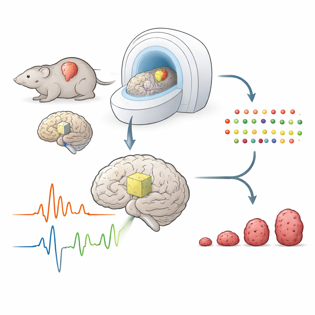
Listening to the chemistry of the brain
Conventional MRI shows where a tumor is, but not what it is doing. Magnetic resonance spectroscopy (MRS) goes further by detecting tiny resonance peaks from different molecules in the brain. Most clinical work so far has focused on the low-frequency "upfield" part of the spectrum, where abundant molecules such as N-acetylaspartate, choline, and lactate are easier to see. The "downfield" region, at higher frequencies, is harder to measure and has often been ignored, even though it may contain signals from crucial molecules involved in energy and protein chemistry. The authors set out to measure both regions at once in rats with and without glioma, using an advanced sequence that preserves fragile downfield signals.
Pairing scans with deep chemical profiling
To understand what the spectral peaks actually represent, the researchers combined in vivo brain spectroscopy with ex vivo untargeted metabolomics. After scanning, they sampled the same brain regions, then used high-resolution liquid chromatography–mass spectrometry to catalog more than 1,600 small molecules. This allowed them to ask, for each MRS peak, which groups of metabolites changed in parallel. They found that tumor tissue showed broad metabolic reprogramming: hundreds of molecules were up- or downregulated compared with normal brain, especially in three major families—nucleotides (the building blocks of DNA, RNA, and energy carriers), lipids, and aromatic compounds known as benzenoids.
Energy currency signals in the downfield region
The most striking discovery was that several downfield peaks rose sharply in glioma and closely tracked markers of the cell’s main energy currency, adenosine triphosphate (ATP), and its breakdown products. Specific downfield resonances near 6.8 and 8.2 parts per million were strongly correlated with metabolites along the ATP pathway, including xanthine, uric acid, and deoxyadenosine. This suggests that those downfield signals serve as indirect, non-invasive fingerprints of heightened nucleotide turnover and energy demand in tumor tissue. In contrast, familiar upfield peaks such as N-acetylaspartate and glutamate mainly reflected loss of normal neurons and shifts in broad metabolic classes, rather than specifically highlighting ATP-related chemistry.
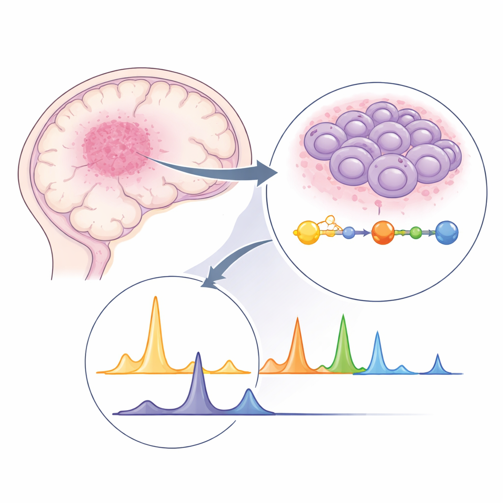
Linking chemistry to how fast tumors grow
Because patients and clinicians care most about whether a tumor is stable or aggressive, the team also asked how the spectral signatures related to tumor size and growth speed. By tracking glioma volume over time in the rats, they calculated each tumor’s growth rate and compared it with its MRS profile. Larger or faster-growing tumors tended to show higher levels of certain metabolites detected in the upfield region, such as lactate and inositol, along with specific downfield peaks tied to nucleotide metabolism. These relationships suggest that the chemical fingerprints captured by MRS do not just reflect static damage, but are tied to the tumor’s dynamic behavior—how quickly it expands and how intensely it burns fuel.
What this means for future brain tumor care
Overall, the study shows that downfield MRS signals, long considered too faint and confusing to use, can act as built-in markers of energy and nucleotide metabolism in glioma. In particular, two peaks around 6.8 and 8.2 parts per million appear to reflect ATP-related pathways that are central to tumor growth. When combined with broader metabolomics, these signals help decode the complex spectrum into meaningful biology and link that biology to how tumors change over time. In the long run, refining these techniques in humans could give doctors a non-invasive way to monitor the metabolic "gears" driving brain tumors, improving diagnosis, risk prediction, and evaluation of therapies that target cancer metabolism.
Citation: Zhu, X., Zhou, K., Cao, Y. et al. Downfield magnetic resonance signals serve as endogenous imaging biomarkers of nucleotide metabolism in glioma. Commun Biol 9, 509 (2026). https://doi.org/10.1038/s42003-026-09780-y
Keywords: glioma, magnetic resonance spectroscopy, nucleotide metabolism, brain tumor imaging, cancer metabolism