Clear Sky Science · en
Distinct origins of human low and high alpha rhythms revealed by simultaneous EEG-SEEG
Why the brain’s quiet waves matter
When you close your eyes and relax, your brain does not fall silent—it hums with gentle electrical rhythms called alpha waves. Anesthesiologists also see strong alpha activity when people are put to sleep for surgery. This study asks a deceptively simple question with big implications: are these alpha waves all the same, or do different kinds of alpha rhythms signal very different brain states, from relaxed wakefulness to loss of consciousness? Understanding the answer could improve how we monitor anesthesia, probe consciousness, and even design new tools for brain health.
Two flavors of a familiar brain wave
Alpha activity is usually treated as one band of rhythms between 8 and 13 cycles per second. The authors show that this range actually hides two distinct rhythms. In awake people with eyes closed, they find a “low alpha” rhythm (about 8–10 cycles per second) strongest at the back of the brain, in regions involved in vision. As doctors increase the dose of the anesthetic propofol and people drift into unconsciousness, this low alpha rhythm fades. At the same time, a “high alpha” rhythm (about 10–13 cycles per second) grows stronger and spreads across the brain, becoming especially prominent once consciousness is lost.
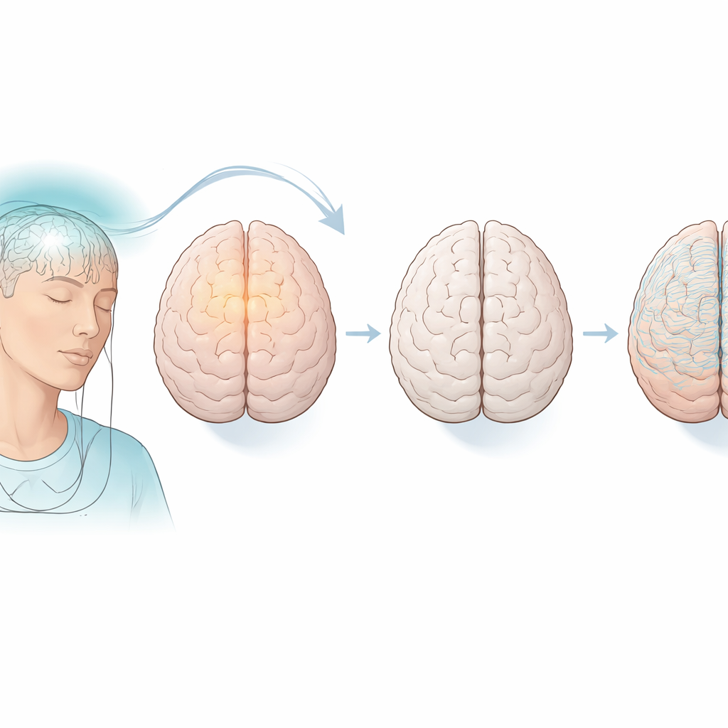
Listening from inside and outside the brain
To uncover these patterns, the team took advantage of a rare opportunity: patients with epilepsy who already had thin depth electrodes implanted in many brain regions for clinical monitoring. While these patients were put under general anesthesia for electrode removal, the researchers recorded signals both from inside the brain (stereo-EEG) and from the scalp (standard EEG). This simultaneous listening from inside and outside allowed them to map where different alpha rhythms were strongest and to check whether signals on the scalp truly reflected activity deep in the brain. They found that, during wakeful rest, low alpha was concentrated in the back of the brain, while under anesthesia high alpha became widespread and more uniform across regions.
Peeling apart true rhythms from background noise
Brain activity at rest is a mix of genuine rhythmic pulses and more irregular background fluctuations. To see which part actually changed with anesthesia, the researchers used a mathematical approach that separates each signal into a “periodic” component (real oscillations like alpha) and an “aperiodic” component (a smooth, noise-like background). They discovered that the dramatic switch from low to high alpha with loss of consciousness was almost entirely due to changes in the true rhythmic component. The background part stayed surprisingly stable. This means the brain is actively reshaping its internal timing, not just changing overall activity levels, when transitioning from a relaxed state to anesthetic unconsciousness.
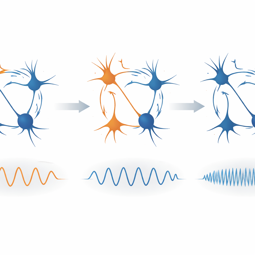
A simple circuit explanation
What could cause low alpha waves in one state and high alpha waves in another? The authors turned to a simple computer model of a local brain circuit made of two players: excitatory cells that tend to activate others, and inhibitory cells that quiet activity down. By slightly increasing the strength of the inhibitory influence—mimicking the action of drugs like propofol that enhance inhibition—they found that the model’s alpha-like rhythm sped up from a lower to a higher frequency, much like the shift seen in their real data. This suggests that tuning the balance between excitation and inhibition in brain circuits can switch the brain between distinct alpha modes linked to wakefulness and unconsciousness.
What this means for sleep, surgery, and brain health
For non-specialists, the key message is that not all alpha waves are created equal. A calm, low alpha rhythm at the back of the head likely reflects a relaxed but awake brain quietly processing less visual information. Under anesthesia, however, a faster, widespread high alpha rhythm may mark a brain whose circuits are tightly held in check by strong inhibition, unable to support conscious experience. Recognizing these distinct alpha signatures could improve how doctors gauge depth of anesthesia, refine theories of consciousness, and support the search for new brain-based markers of conditions such as dementia, depression, or attention disorders. In short, the familiar alpha wave turns out to be a more nuanced window into the brain’s shifting states than previously thought.
Citation: Wang, R., Jiang, S., Cai, Q. et al. Distinct origins of human low and high alpha rhythms revealed by simultaneous EEG-SEEG. Commun Biol 9, 503 (2026). https://doi.org/10.1038/s42003-026-09769-7
Keywords: alpha brain waves, anesthesia, consciousness, EEG, neural oscillations