Clear Sky Science · en
Scalable and multimodal brain angiogenesis and blood-brain barrier genetics by somatic mutagenesis
Why Protecting the Brain’s Border Matters
The brain sits behind a microscopic security fence called the blood–brain barrier, which carefully controls what can pass from the bloodstream into our most sensitive organ. When this barrier fails, it can contribute to stroke, dementia, epilepsy, and other neurological diseases—but it also blocks many promising drugs from reaching the brain. This study introduces a fast, scalable way to test which genes keep this barrier healthy or make it leak, using a combined approach in zebrafish and mice. By speeding up gene discovery, the work opens new paths toward treating brain disorders and safely delivering medicines into the brain.
Two Tiny Animals, One Big Question
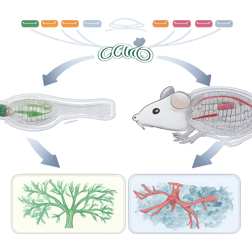
The researchers set out to build a practical testing platform rather than focusing on a single disease. Their goal was to learn, in weeks instead of years, which genes control the growth of brain blood vessels and the tightness of the blood–brain barrier. No single animal model is ideal for every stage of this system: early vessel growth in mammals is hidden deep inside the embryo, while the adult brain vasculature in tiny fish is hard to probe in detail. The team therefore combined the strengths of two well-established laboratory animals. Transparent zebrafish embryos let scientists watch new brain vessels form in real time, while adult mice provide a realistic setting to test how well the mature barrier blocks unwanted molecules.
Watching Brain Vessels Grow in Living Fish
To study how brain vessels first form, the team used zebrafish embryos whose blood vessels glow under the microscope. They injected freshly fertilized eggs with molecular tools that cut specific genes in many cells at once, creating what are called somatic mutants. Within a day or so, they could directly count fine vessel sprouts in the hindbrain and compare them with normal patterns in the fish’s trunk, which served as a control. By targeting known regulators of vessel growth and barrier function, including genes involved in blood–brain barrier seals, signaling, and nutrient transport, they showed that their fish assay reliably reproduced expected defects. Some genes caused clear reductions in brain vessel branching, while others left early vessel growth unchanged, revealing which ones matter at this stage.
Stress-Testing the Barrier in Adult Mice
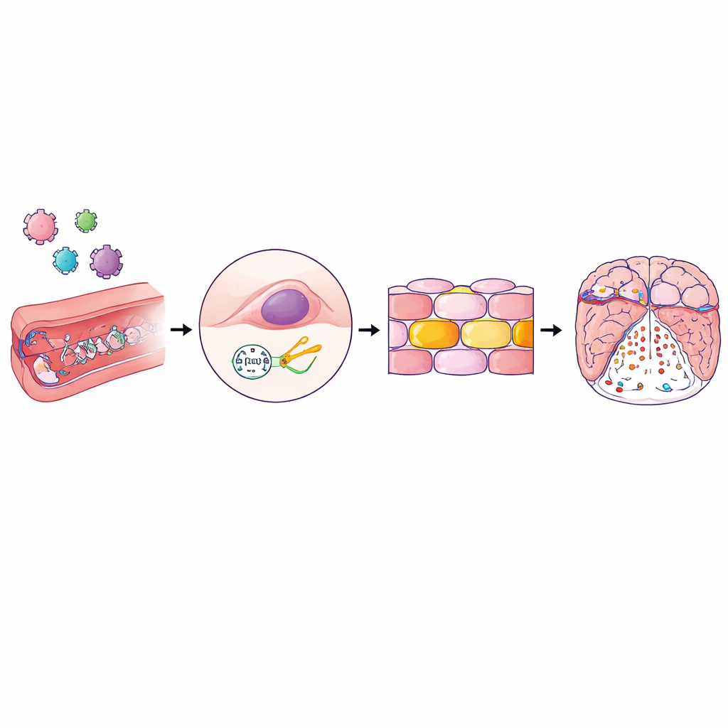
Brain vessel growth is only the first chapter; the barrier must then stay tight for a lifetime. To probe this long-term gatekeeping, the researchers turned to mice engineered so that their brain vessel cells can express the CRISPR cutting protein. They packaged sets of guide molecules into specially designed viral particles that home to brain blood vessels after a simple injection into the bloodstream. Once inside the vessel-lining cells, these guides direct CRISPR to snip selected genes in a patchwork, or mosaic, fashion. The team then monitored the animals for seizure-like behavior, a sensitive sign of neurovascular stress, and injected a small fluorescent dye that normally cannot cross an intact barrier. By measuring how much dye leaked into brain tissue and visualizing its spread in brain slices, they could quickly tell which gene disruptions weakened the barrier.
Fast Answers from a Gene-by-Gene Test Drive
Using this two-species platform, the authors retested a set of genes already known to influence brain vessels and barrier integrity, including claudin-5 (a key component of the barrier’s tight seals), β-catenin (a central signaling hub), a glucose transporter, a protease, and a regulator of inflammatory signaling called Nemo. Their zebrafish assay confirmed that certain genes are specifically needed for brain vessel sprouting, while others are not. In mice, disrupting barrier seal genes or core signaling components caused seizures and allowed the fluorescent dye to seep into the brain, mirroring earlier, slower studies using traditional breeding. Nemo, in contrast, was shown to be crucial for barrier protection in adult mice but dispensable for initial vessel growth in fish. Crucially, each full round of testing—from designing CRISPR guides to reading out fish and mouse results—could be completed in about six weeks and multiplexed across several genes in parallel.
What This Means for Brain Health and Future Therapies
For non-specialists, the key message is that this study delivers a practical “test bench” for the genes that build and guard the brain’s vascular border. Instead of spending months or years generating traditional mutant animals one by one, researchers can now disrupt candidate genes rapidly in zebrafish and mice, watch how brain vessels grow, and measure how leaky or tight the barrier becomes. While the method will not uncover every gene involved, its speed and flexibility make it well suited to exploring large gene lists from human genetics or brain disease studies. Over time, mapping this genetic control system could reveal new drug targets to repair a failing barrier or to open it temporarily and safely, bringing us closer to better treatments for conditions ranging from epilepsy to vascular dementia.
Citation: Panji, J.M., Germano, R.F.V., America, M. et al. Scalable and multimodal brain angiogenesis and blood-brain barrier genetics by somatic mutagenesis. Commun Biol 9, 479 (2026). https://doi.org/10.1038/s42003-026-09747-z
Keywords: blood-brain barrier, brain angiogenesis, CRISPR screening, zebrafish and mouse models, neurovascular genetics